

RESIDUES IN HONEY OF OXYTETRACYCLINE AND TYLOSIN USED FOR THE CONTROL OF AMERICAN FOULBROOD DISEASE IN SLOVAKIA
tekst: Juraj Toporčák, Jozef Nagy, Jozef Sokol
Pszczelnicze Zeszyty Naukowe, Rok XXXIX, 1995 — Juraj Toporčák, Jozef Nagy, Jozef Sokol
strona 113PSZCZELNICZE ZESZYTY NAUKOWE Rok XXXIX, Nr 1 1995
Juraj Toporčák, Jozef Nagy, Jozef Sokol
University of Veterinary Medicine in Košice, Komenského 73, 041 81 Košice, SLOVAK REPUBLIK
Summary
The sulfonamides were used to control bacterial diseases. But after new information (Turell, Gliński, Otte, Drobnikova) and scientific survey about resistency of B.larvae on sulfonamides we changed it for oxytetracyclin and tylosin. Oxytetracyclin is the most frequently used against American Foulbrood in the world. Tylosin was firstly tested in USA by Hitchcock on bees and later by Drobnikova. Machova firstly paid attention on tylosin residues in honey by using microbiological method.
In our testings we followed the residues of oxytetracyclin and tylosin in 3 different doses on bee colonies. The residues of antibiotics were followed in week intervals by microbiological method. From those results can be said that tylosin used for control of AFB was recorded in honey longer period then oxytetracyclin but its advantage is that tylosin is used only in veterinary medicine.
Keywords: honeybee, honey, oxytetracyclin, tylosin, residues.
INTRODUCTION
American foulbrood (AFB) is one of the most dreaded bee diseases in the world. It is a highly contagious infectious disease caused by the spore forming bacterium Bacillus larvae and is found almost worldwide. Bacillus larvae forms oval very resistant endospores and therefore according to Veterinary State Laws all positive colonies in an apiary have to be destroyed by burning. The negative colonies in the apiary are preventively controlled by drugs.
Sodium sulfathiazole was used for many years but after the resistance of Bacillus larvae to sulphonamides had been stated (Turrel 1974; Glinski and Rzedzicki 1977; Otte 1972; Drobnikova 1983, 1985), this substance was replaced by oxytetracycline and later tylosin on the basis of the results of Drobnikova et al. (1987). In the USA Hitchcock et al. (1970) were the first to test tylosin against AFB in bees. The advantage of tylosin is that it is used only in veterinary practice. Its disadvantage is the risk of occurrence of tylosin residues in the honey and syrup stored by the honeybees; this occurrence depends on drug administration and the ability of macrolide antibiotics to quickly cause chromosomal resistance of
strona 114bacteria. Tetracycline and oxytetracycline were used in the control and prevention of AFB by Gochnauer and Bland (1974), Shimanuki (1978) and Bailey (1981), and chlortetracycline was tested by Peng et al. (1992).
Similarly the same drugs are used to treat another bacterial disease – European foulbrood.
The Disc-assay system for detection of antibiotics in honey was used by Gochnauer and Bland (1974), Wilson (1974), Bentler and Frese (1981), Lehnert and Shimanuki (1981) and Roth et al. (1986). The HPLC method was used by Jurgens (1981), Serra i Bonvehi and Gomez Pajuelo (1983, 1984), Sporns et al. (1986), Matsuka and Nakamura (1990), Galeano Diaz et al. (1990), Argauer and Moats (1991), Glinsky et al. (1991) and Nakaya et al. (1991).
MATERIAL AND METHODS
Honey bees. In the, 36 honey bee colonies (for every test dose 6 colonies) kept at the University of Veterinary Medicine in Košice were used. One colony consisted of 4 brood and 2 empty combs.
Antibiotics. In the test Oxymykoin pulv. solub. a.u.v. Spofa with 5,5 g oxytetracycline hydrochloride in 100 g powder and a macrolide antibiotic, Tylan Elanco 200, inj. a.u.v. with 200 mg tylosin in 1 ml of solution were used.
The drugs (1 application) were fed in sugar syrup (2 parts of sugar to 1 part of water) during the spring period.
Antibiotic activity was detected 1 week after feeding.
Doses:
Oxymykoin pulv. – 3 groups per 6 colonies:
- – 2.5 g Oxymykoin (0.137 g OTC)/ colony
- – 5.0 g Oxymykoin (0.275 g OTC)/ colony
- – 10.0 g Oxymykoin (0.550 g OTC)/ colony
Tylan Elanco 200 inj. – 3 groups per 6 colonies:
- – 0.4 g tylosin (2 ml Tylan + 0.8 ml 96% ethanol)/ colony
- – 0.8 g tylosin (4 ml Tylan + 1.6 ml 96% ethanol)/ colony
- – 1.6 g tylosin (8 ml Tylan + 3.2 ml 96% ethanol)/ colony
Disc-assay method. A honey sample of 1 g was accurately weighed, heated to 70 oC for 15 minutes. Filter paper discs (o 12 mm) were dipped in to the honey. and then placed on the surface of the inoculated medium. Two test
strona 115organisms, Bacillus stearothermophilus, var. calidolactis C 953 and Bacillus subtilis BGA (pH of medium 6.0; 7.2; 8.0) were evaluated for their sensitivity to OTC and tylosin.
HPLC method. OTC was obtained from Sigma Chemical, acetonitrile, methanol and HPLC grade were obtained from Merck, oxalic acid was obtained from Lachema and [cellulose] membrane filters were obtained from Hewlett Packard.
The samples were tested by the HP 1050 S Hewlett Packard, liquid chromatograph, consisting of quarter pump, autosampler, „VU – VIS detector and 3396” Integrator.
The samples were prepared according to the method of Galeano Diaz et al. (1990) and analyzed by HPLC isocratic mode on the Spherisorb ODS2 125 mm column using the acetonotril-methanol-0.01 M oxalic acid 25:15:60 at a flow rate of 0.7 ml/min. UV detection was carried out at 360 nm.
RESULTS AND DISCUSSION
As to the test organisms employed, detection of OTC residues by the disc-assay system was based on the methods used under the conditions of this country. The presence of the given antibiotic (Fig. 1) became evident by zones of inhibition measuring 9-16 mm in diameter when Bacillus stearothermophilus was used. Gradually an abrupt decrease to 2-6 mm occurred. However, the above test microorganism is known for its high sensitivity to natural inhibitors, great amounts of which are particularly present in honey.
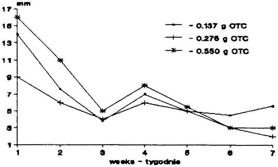
Fig.1. The mean zone diameters of the honey samples with OTC tested on Bacillus stearothermophilus
Ryc.1. Przeciętna średnica strefy zahamowania wzrostu Bacillus stearothermophilus w próbkach miodu z OTC
Much smaller zones of inhibition were measured when the samples were tested by Bacillus subtilis (Fig. 2 – mean values). The latter revealed maximum
strona 116sensitivity when pH of the medium was 6.0. At the first sampling the maximum zones of inhibition were 6 and 2.8 mm for the groups with the highest and lowest dose. A gradual decrease of the inhibition zones followed; in the last week no inhibition zones were observed in Groups 1 and 2 and a zone less than 1 mm was detected in the group receiving maximum concentrations.
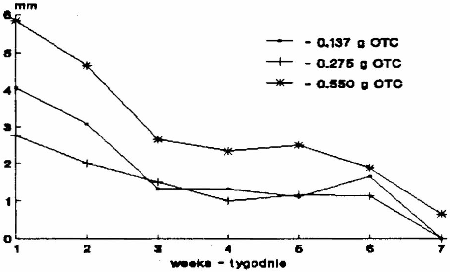
Fig.2. The mean zone diameters of the honey samples with OTC tested on Bacillus subtilis
Ryc.2. Przeciętna średnica strefy zahamowania wzrostu B.subtilis w próbkach miodu z OTC
Some authors have demonstrated OTC in honey after the elimination of natural inhibitors by diluting samples in distilled water and subsequent heat-processing using catalase, peroxidase, phosphate buffer at pH 7.0 (Roth et al., 1986) and the test strain Bacillus cereus. Wilson (1974) employed Bacillus cereus var. mycoides (ATCC 11778) after having treated the samples with phosphate buffer (pH 4.5). He reported a minimum sensitivity of 0.25 ppm (μg/g) for all samples.
The results obtained by observing tylosin residues after feeding tylosin to honey bees are summarized in Fig. 3 and 4.
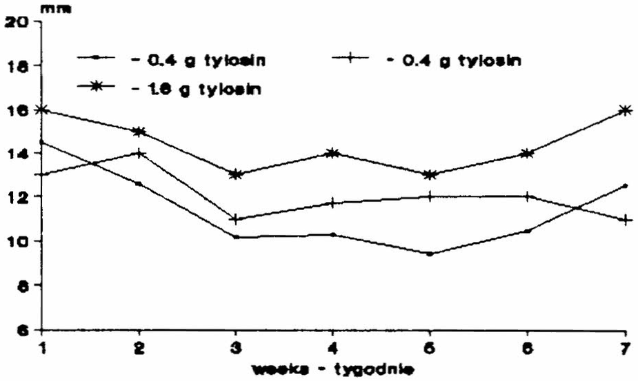
Fig.3. The mean zone diameters of the honey samples with tylosin tested on Bacillus stearothermphilus
Ryc.3. Przeciętna średnica strefy zahamowania wzrostu Bacillus stearothermphilus w próbkach miodu z tylozyną
strona 117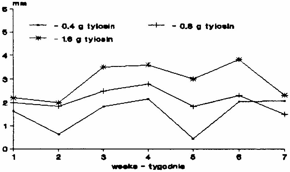
Fig.4. The mean zone diameters of the honey samples tylosin tested on Bacillus subtilis
Ryc.4. Przeciętna średnica strefy zahamowania wzrostu Bacillus subtilis w próbkach miodu z tylozyną
Honey samples examined for tylosin residues by Bacillus stearothermophilus as the test organism inhibited the growth of the latter similar to OTC, since the maximum mean inhibition zone in the group receiving the maximum concentration was 16 mm in diameter. In contrast to OTC observations tylosin levels were present at minimum fluctuation up to the last sampling, and the size of the inhibitions zones was similar to that seen at the start of the examination.
The course of examination using Bacillus subtilis resembles the smaller inhibition zones caused by the different sensitivity of the strains under comparison (most sensitive – Bacillus subtilis at pH of the medium 8.0).
Generally it can be stated that comparison of Bacillus subtilis and B.stearothermophilus revealed a low sensitivity of the former to tylosin. The diameter of the inhibition zones only surpassed 3 mm in the case of the maximum concentration used.
As to HPLC, OTC feeding lead to different OTC levels in the honey according to the concentrations used (Fig. 5). The maximum OTC level presented 20.54 mg.kg-1 in the group with the maximum concentration. In the following weeks a pronounced decrease of OTC levels was observed in the samples examined. In the last week (week 8), concentrations at the detection limit of the given method (0.17 mg.kg-1) were measured in Group 1. In Group 2 OTC levels reached 0.497 mg.kg-1 and in the group with the maximum doses fedthe mean values decreased below 1.0 mg.kg-1. Gilliam and Argauer (1981) reported OTC to be degraded in honey 4 weeks after the withdrawal of icing sugar or syrup treatment of bees. Similarly, in our experiment it was after 4 weeks that the values observed in the two groups receiving less OTC approached the detection limit of the method. Gliński et al. (1991) stated Terramycine degradation to occur within 6-8 weeks.
strona 118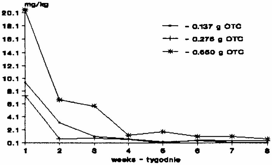
Fig.5. The levels of OTC in the honey samples determined by HPLC
Ryc.5. Poziom OTC w próbkach miodu, określony metodą HPLC
REFERENCES
Argauer R. J., Moats W. A. (1991) – Degradation of oxytetracycline in honey as measured by fluorescence and liquid chromatographic assays. Apidologie, 22:109-115.
Bailey L. (1981) – Honey Bee Pathology. Academic Press, London.
Bentler V. W., Frese E. (1981) – Mikrobielle Beschaffenheit und Ruckstandsuntersuchung von Bienenhonig. Archiv. Lebensmeittelhyg, 32:130-135.
Drobnikova V. (1983) – Studium bacteriofagu a plasmidu Bacillus larvae. Research Institute of Beekeeping, Dol. Final Report.
Drobnikova V. (1985) – Vyzkum tlumeni bakterialnich nakaz vcel. Research Institute of Beekeeping, Dol. Final Report.
Drobnikova V., Pytelova J., Mala J. (1987) – Vyzkum infekcnich nakaz vcel. Resesarch Institute of Beekeeping, Dol. Final Report.
Galeano Diaz T., Guiberteau Cabanillas A., Salinas F. (1990) – Rapid determination of sulfathiazole, oxytetracycline and tetracycline in honey by high-performance liquid chromatography. Analytical letters, 23(4):607-616.
Gilliam M., Argauer R. J. (1981) – Oxytetracycline residues in surplus honey, brood nest honey, and larvae after medication of colonies of honey bees, Apis mellifera, with antibiotic extender patties, sugar dusts, and syrup sprays. Environ. Entomol., 10:479-482.
Gliński Z., Rzedzicki J. (1977) – Evaluation of the sensitivity of Bacillus larvae to antibiotics and sulfonamids isolated in Poland in 1962-1971. Pol. Arch. weter., 20:9-16.
Gliński Z., Kauko L., Gacek G. (1991) – Badania nad pozostałością oksytetracykliny w miodach konsumpcyjnych z pasiek leczonych przeciwko zgnilcowi złośliwemu. Med. Wet., 47(12):534-536.
Gochnauer T. A., Bland S. E. (1974) – Persistence of tetracycline activity in medicated syrup stored in honeybee colonies in late spring. J. Apic. Res., 13:153-159.
Hitchcock J. D., Moffett J. O., Lackett J. J., Elliott J. R. (1970) – Tylosin for control of American foulbrood disease in honey bees. J. econ. Entom., 63:204-207.
Jurgens U. (1981) – Zur hochdruckflussigchromatographischen analyse von arzneimittelruckstanden in honig. Z. Lebensm. Unters. Forsch., 173: 356-358.
Lehnert T., Shimanuki H. (1981) – Oxytetracycline residues in honey following three
strona 119different methods of administering the antibiotic. Apidologie, 12: 133-136.
Machova M., Drobnikova V., Titera D., Vejdovska A., Kristanova D., Henzl S. (1992) – Preventive use of tylosin in the colonies endangered by the foulbrood and residues of tylosin in honey and syrup stored by honeybees. Veterinarni Medicina, 37:119-125.
Matsuka M., Nakamura J. (1990) – Oxytetracycline residues in honey and royal jelly. J. Apic. Res., 29(2):112-117.
Nakaya K., Kobayashi Y., Tanahashi N. (1991) – Fluorometric determination of tetracyclines in honey by high performance liquid chromatography. J. Food Hyg. Soc. Japan, 32:43-47.
Otte E. (1972) – Die Empfinlichkeit von Bacillus larvae stammen gegen chemoterapeutica. In: XXIII. Int. Bienenzucht kongress, Apimondia, Bucharest: 462-463.
Peng Ch. Y-S., Mussen E., Fong A., Montague M. A., Tyler T. (1992) – Effects of chlortetracycline of honey bee worker larvae reared in vitro. J. Apic. Res.,60: 127-133.
Roth L. A., Kwan S., Sporns P. (1986) – Use of a disc-assay system to detect oxytetracycline residues in honey. J. Fd. Protect., 49:436-441.
Shimanuki H. (1978) – Bacteria, In: R.A. Morse,[ed.], Honey Bee Pests, Predators, and Diseases. Cornell University Press, Ithaca, New York.
Serra i Bonvehi J., Gomez Pajuelo A. (1983) – Contaminacion de la miel residuos medicamentosos (O.T.C. y sulfamidas) procedentes de la medicacion preventiva a las colonias de abejas. Apiacta, 18(4):109-110.
Serra i Bonvehi J., Gomez Pajuelo A. (1984) – Contamination du miel par des residus medicamenteux (oxytetracycline et sulfamides) provenant de traitements preventifs administres aux colonies d’abeilles. Bul. Tech. Apic., 11(4):203-206.
Sporns P., Kwan S., Roth L. A. (1986) – HPLC analysis of oxytetracycline residues in honey. J. Food Protection, 49:383-388.
Turell M. J. (1974) – A history of the use of drugs in the prevention and cure of American foulbrood. Am. Bee J., 114:13.
Wilson W. T. (1974) – Residues of oxytetracycline in honey stored by Apis mellifera. Environ. Entomol., 3:674-676.
POZOSTAŁOŚCI W MIODZIE OKSYTETRACYKLINY I TYLOZYNY – ANTYBIOTYKÓW STOSOWANYCH DO ZWALCZANIA ZGNILCA ZŁOŚLIWEGO (AFB) NA SŁOWACJI
J. Toporčák, J. Nagy, J. Sokol
Streszczenie
Sulfonamidy od wielu lat stosowano do leczenia chorób zakaźnych. Nowe doniesienia naukowe (Turell, Gliński, Otte, Drobnikova) na temat występowania szczepów B.larvae opornych na podawane sulfonamidy, wpłynęły na coraz powszechniejsze stosowanie antybiotyków w terapii zgnilca złośliwego (AFB).
Najszersze zastosowanie spośród antybiotyków znalazła oksytetracyklina. Antybiotyk makrolidowy – tylozynę, do walki ze zgnilcem złośliwym zastosował po raz pierwszy Hitchcock, a następnie Drobnikova. Problem pozostałości tylozyny w miodzie poruszyła Machova, która w swoich badaniach stosowała metody mikrobiologiczne.
strona 120Nasze badania miały na celu wykrycie pozostałości oksytetracykliny i tylozyny w miodzie. Obydwa antybiotyki zostały podane rodzinom pszczelim w trzech różnych dawkach. Obecność antybiotyków w miodzie określano metodą mikrobiologiczną, w cotygodniowych odstępach. Na podstawie otrzymanych wyników stwierdzono występowanie pozostałości tylozyny w miodzie przez dłuższy okres czasu aniżeli oksytetracykliny. Zaletą tylozyny jest jedynie fakt, że jest ona stosowana wyłącznie w medycynie weterynaryjnej.
Słowa kluczowe: pszczoła miodna, miód, oksytetracyklina, tylozyna, pozostałości.
Errata — błędy druku znalezione podczas digitalizacji
- s. 113: „REPUBLIK” → REPUBLIC
- s. 113, streszczenie: „period then” → period than
- s. 113: „Turrel” → Turell
- s. 114: „Glinsky” → Gliński
- s. 115: „VU – VIS” → UV – VIS
- s. 115: „acetonotril” → acetonitrile
- s. 116, ryc. 3, 2×: „stearothermphilus” → stearothermophilus
- s. 116, ryc. 3, legenda, druga linia: „0.4 g tylosin” → 0.8 g tylosin
- s. 117: „inhibitions zones” → inhibition zones
- s. 117: „fedthe” → fed the
- s. 118, piśmiennictwo: „Lebensmeittelhyg” → Lebensmittelhyg
- s. 118, piśmiennictwo: „Resesarch” → Research
- s. 119, piśmiennictwo: „Empfinlichkeit” → Empfindlichkeit