

LONG QUEEN CELLS DISEASE
tekst: Jerzy Woyke, Jerzy Bobrzecki
Pszczelnicze Zeszyty Naukowe, Rok XXII, 1978 — J. Woyke, J. Bobrzecki
strona 59PSZCZELNICZE ZESZYTY NAUKOWE ROK XXII 1978
J. Woyke
Institut for Biavl, Norges Landbrugshøgskole, Asker, Norway *
J. Bobrzecki
Norges Birøkterlag, Billingstad, Norway **
INTRODUCTION
There appeared in Norway in 1972 a disease which effected the queen rearing so seriously, that it was quite impossible to rear any queen.
Mr. O. Rosenberg, head of the Bee Station, describes the history as follows: ”We started our grafting about May 29th. The queens developed normally and emerged in our incubator to the expected date. On the 4th June Dr. J. Bobrzecki grafted about 100 queen cells which developed into very long cells. We did not understand why they were abnormal. I have to insert that the climate in June was very unfavorable to queen breeding — rain, low temperature and no honey flow, and in particular a serious lack of pollen. All of this together has of course favoured the development of fungi — chalk brood which has caused an epidemic in Norway for the last six — seven years. In one colony placed in one of our outyards we also found sack brood.
In about 4 weeks after the first group of queen cells, Dr. Bobrzecki had grafted about 2000 cells which had given about 1000 queen cells, but not a single normal queen emerged. In the beginning, the first two weeks, we noticed that the cells were not sealed at the right time and
Present adress
- Zakład Owadów Użytkowych, SGGW — Akademia Rolnicza, Warszawa, Ursynów, Polska
** Zakład Pszczelarstwa, Akademia Rolniczo-Techniczna, Olsztyn, Kortowo, Polska
strona 60many of the cells were not sealed at all. As we ordinarily are short of royal jelly we took the jelly from the open cells which sometimes were up to 5 cm long.
So after a certain period of time we had very much royal jelly which was used in the grafting. As the Institute for Beekeeping is our neighbour, 6 km away, and they had no royal jelly for their own grafting they asked us for a quantity. They grafted 50—60 cells with this jelly and after some days they got the same results — long cells with dead larvae. Unfortunately the pathology gives very limited information about chalk brood. It is a disease considered to be rather harmless. But if we are right in our assumptions about chalk brood it is clear that it can be a serious problem”.
The pathology and epidemiology of the described brood diseases concerns mostly worker brood and sometimes also drone brood. We therefore undertook investigation to describe it in the queen brood.
MATERIAL AND METHODS
The investigation was conducted in the summer of 1972 in the Beekeeping Station of the Norwegian Beekeeper Association, in the Bee Culture Institute of the Norwegian Agricultural College and finished in Poland.
Queen cells taken from colonies producing abnormal, long cells were mostly investigated. But normal queen cells reared by other colonies were also used as a control. There were measured 91 queen cells taken from colonies producing the long cells, and 19 normal queen cells. Sealed queen cells were put into an incubator at 34—35°C. Brood in the cells was examined at various periods before and after sealing.
Cocoons of normal and long queens cells were investigated when the queens emerged or at least 20 days after grafting the larvae. The queen cells were diped into worm xylen which dissolved the wax.
It is very popular in Scandinavia to use old cells for grafting in which a queen developed already. The cells are shortened and given to a rearing colony, where they are cleaned and rebuilt into queen cups. Then they are used for grafting again. To detect the ways of infection both kinds of cups were used, those being rebuilt from the used cells and new cups. Each cup was supplied with a droplet of food. Three kinds of it were applied, either royal jelly collected from the long queen cells, jelly from normal queen cells or honey. So six combinations were made. Then 667 youngest larvae were grafted. About 10 coups with larvae of one combination were attached to one nursery bar. Two to three bars with different combinations of cells and food were placed in one nursery frame and then given to a queenless rearing colony. The number of accepted larvae was checked the next day and the kind of resulting queen cells was examined 5 days after grafting.
strona 61Attempts were undertaken to infect the queen larvae with chalk-brood disease. For this purpose five chalked larvae covered with mycelium and spores were crushed in 1 cm3 of water. Next larvae accepted for one day were moved out of queen cells. The jelly in those cells was mixed with the water suspension of the infected larvae and then new larvae one day old were grafted again. Two repetitions were made. The first time all 16 accepted cells were regrafted. The second time, out of 13 accepted cells only 7 were regrafted and the remaining 6 were left as a control group.
In a second attempt one chalked larva was crushed in roval jelly diluted with a little water. Then the jelly with the larval crush was kept for one day in a incubator at 34°C and for two days at room temperature. This jelly was used for grafting 20 larvae into new cups. Results of both attempts were checked the next day, then 5 days after giving the larvae to the queenless colony and again when the queens emerged.
RESULTS
LENGTH OF QUEEN CELLS
Measurement of a control group of 19 sealed normal queen cells in a helthy colony showed, that they were from 23 mm. to 31 mm. long, with an avg. of 26.5 ±0.55 mm. and standard deviation (S) of 2.4 mm.
Fig. 1 shows the abnormal queen cells, taken from colonies producing long cells.
Some of the abnormal queen cells were of normal size but they were ended with duller lids. The longer cells were often contracted in their lower part. The longest looked like those prolongued ones containing drone larvae. They are known to any queen breeder. Measurements of 91 cells, from colonies producing long queen cells, showed that they were from 23 mm. to 60 mm. long, with an avg. of 32.6 ±0.65 mm. S = 6.17 mm. So they averaged 6.1 mm. longer than the normal queen cells. The distribution of length presented in diagram, (Fig. 2) showed, that 86% of cells were collected in a range from 24 mm. to 37 mm. Single cells only we were found in classes outside this range.
CONTENTS OF THE QUEEN CELLS
Live larvae were found in unsealed and just sealed long queen cells. The weight of 5 larvae from just sealed cells ranged from 155 mg. to 203 mm. and the avg. was 173 mg. The same number of larvae taken in the same stage from normal queen cells weighed from 154 mg. to 221 mg. and the avg. was 179 mg. Thus larvae from the prolonged cells were a little lighter but, no drastic underdevelopment was found at this time.
strona 62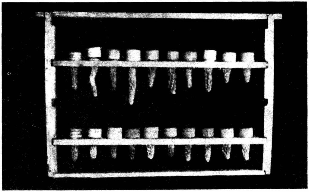
Fig. 1. Long queen cells. Two cells on the right side of each bar are normal. On the upper bar are long cells, on the lower bar are short and normal length cells ended with dull lids.
Ryc. 1. Długie mateczniki. Dwa mateczniki z prawej strony każdej listewki sa normalne. Na górnej listewce pokazano mateczniki przedłużone. Na dolnej mateczniki nienormalne o skróconej lub normalnej długości lecz zakończone tępym wieczkiem.
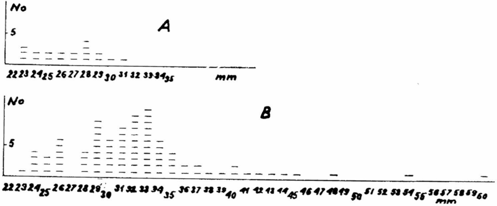
Fig. 2. Length distribution of queen cells. A — normal, B — abnormal and long cells.
Ryc. 2. Rozdział długości mateczników. A — normalnych, B — nie normalnych i przedłużonych.
Observation of larvae through a hole made in the queen cells placed in an incubator showed that the larvae were moving inside. They spun the cocoon and ate the royal jelly adhered to the base of the cells. But the movements of the larvae in the long cells were less vigorious than of those in the normal ones.
strona 63Healthy larvae are pupated already the fourth day after the sealing of the cells. Examination of 37 long cells, 5 days after sealing, showed in 7 of them, larvae overgrown with mycelium. They resembled the chalk brood. Brood in the remaining 30 cells was also not pupated. It was in the prepupa stage. A transparent fluid was accumulated beneath the skin. The prepupae dried after a few days and changed the colour to brown and later to black. The characters described resembled sac brood disease.
But the sac was not as typical, as that of infected worker larvae in brood combs. Many larvae died in earlier stage. Perhaps being weaker they could not reach the food in the base of the queen cell after it was sealed, and the larvae starved.
Some characters, like loss of plumpness and transparent skinn resembled early stages of European faulbrood. But the prepupae did not become white, cream and ropy and did not decompose. The undersized adults could eventually develope from larvae infected by Streptococus pluton. Probably the feeding and development conditions different in worker and queen cells could change the characters typical for infected worker larvae or could mix characters typical for different diseases.
Several other series of long queen cells were left for a longer period in an incubator. The percentage of cells with chalk brood did not increase. In a few cells, brown or black prepupae were covered with mold. But it seems this was a secondary contamination.
Examination of cocoons released from the wax by xylen showed, that these from the normal cells were distinctly thicker than those from the prolonged cells. Evidently the larvae were weaker or did not produce as much spining excretion as the healthy larvae.
WAYS OF INFECTION
After the larvae were grafted on various food in two types of cups four kinds of queen cells were found five days after grafting: normal sealed queen cells, prolonged sealed cells, prolonged unsealed and empty cells without larvae. Empty cells are not presented in tables, unsealed cells should be counted as abnormal ones.
After the data were collected, two very distinct groups appeared. One concerned cells sealed before July 6th and the other those sealed after that date. For better analysis the results are presented in two tables No. 1 and No. 2.
Table No. 1 shows that altogether more larvae were accepted on royal jelly (73—85%) than on the honey (42—58%). Concerning the kind of cups, out of all 204 larvae grafted in used cups on various food 70% were accepted whereas out of 324 grafted into new cups 73% were reared. So there was no significant difference found in the acceptance of larvae grafted into used or new cups.
strona 64Table 1 No. of normal and abnormal queen after grafting the larvae into two kinds of cups on three different diets (before July 6) Tabela 1. Liczba normalnych i nienormalnych mateczników po przełożeniu larw do dwu grup miseczek matecznikowych na trzy rodzaje pokarmu (przed 6 lipca)
| Used cups Miseczki używane: Grafted larvae Przełożono larw | Used cups Miseczki używane: Accepted next day Przyjęto | Used cups Miseczki używane: Queen cells after 5 days Mateczniki po 5 dniach: long długie | Used cups Miseczki używane: Queen cells after 5 days Mateczniki po 5 dniach: unsealed niezakryte | Used cups Miseczki używane: Queen cells after 5 days Mateczniki po 5 dniach: normal normalne | New cups Miseczki nowe: Grafted larvae Przełożono larw | New cups Miseczki nowe: Accepted next day Przyjęto | New cups Miseczki nowe: Queen cells after 5 days Mateczniki po 5 dniach: long długie | New cups Miseczki nowe: Queen cells after 5 days Mateczniki po 5 dniach: unsealed niezakryte | New cups Miseczki nowe: Queen cells after 5 days Mateczniki po 5 dniach: normal normalne |
|---|---|---|---|---|---|---|---|---|---|
| Royal jelly from long queen cells Mleczko z długich mateczników | |||||||||
| 10 | 10 | 7 | 20 | 19 | 15 | 1 | 1 | ||
| 10 | 10 | 4 | 3 | 10 | 7 | 3 | 2 | ||
| 11 | 6 | 1 | 2 | 10 | 9 | 2 | 5 | ||
| 20 | 15 | 1 | 7 | 10 | 9 | 8 | |||
| 10 | 10 | 5 | 10 | 7 | 6 | ||||
| 61 | 51 | 18 | 5 | 7 | 61 | 51 | 34 | 3 | 6 |
| % | 85 | 30 | 8 | 12 | % | 85 | 57 | 5 | 10 |
| Fresh royal jelly from normal queen cells Świeże mleczko z normalnych mateczników | |||||||||
| 10 | 5 | 2 | 10 | 1 | 1 | ||||
| 10 | 10 | 9 | 10 | 9 | 8 | ||||
| 10 | 6 | 3 | 1 | 11 | 7 | 1 | 6 | ||
| 10 | 7 | 2 | 20 | 15 | 15 | ||||
| 20 | 19 | 2 | 16 | 21 | 19 | 1 | 5 | 13 | |
| 10 | 10 | 8 | 2 | 21 | 19 | 19 | |||
| 11 | 8 | 3 | 5 | 131 | 94 | 3 | 1 | 82 | |
| 81 | 65 | 27 | 2 | 24 | 224 | 164 | 5 | 6 | 144 |
| % | 80 | 33 | 2 | 30 | % | 73 | 2 | 3 | 64 |
| Honey Miód | |||||||||
| 10 | 2 | 2 | 10 | 10 | 3 | ||||
| 10 | 1 | 1 | 10 | 1 | 1 | ||||
| 10 | 4 | 4 | 10 | 3 | 3 | ||||
| 22 | 12 | 11 | 10 | 9 | 8 | ||||
| 10 | 7 | 6 | |||||||
| 62 | 26 | 5 | — | 19 | 40 | 23 | — | — | 15 |
| % | 42 | 8 | 31 | % | 58 | 38 | |||
| Total Razem | |||||||||
| 204 | 142 | 324 | 238 | ||||||
| % | 69.6 | % | 73.4 |
Within the group supplied with royal jelly taken from long queen cells (Table 1), 57% of long queen cells were built on new cups and 30% on the used ones.
strona 65The lower percentage of long cells built on used cups did not show lower infection. Altogether larvae were found the fifth day in 50% of used cells and in 72% of the new ones. There can be concluded, that the infection was so high in used cups that the larvae were lost before being sealed. The percentage of normal queen cells build or both kinds of cups was similar but very low (10—12%).
Grafting the larvae on royal jelly taken from normal cells (Tab. 1) increased the percentage of normal queen cells built on the used cups up to 30%. But still similar percentage of abnormal cells (33%) was produced on the used cups. Contrary to this, twice as many normal cells were build on new cups (Tab. 1) 64% and only a few 2% abnormal appeared. Grafting larvae on honey into new cups resulted in all normal queen cells but in low percentage (38%). Few abnormal cells were found among those built on used cups (Tab. 1).
All data presented shows, that the larvae could be infected as well by the used cups as by royal jelly collected from long queen cells. After new cups were supplied with fresh normal jelly, the infection was almost eliminated. Single exceptions could be caused by infection from the contaminated cells and food in other bars of the same frame.
Table 2 shows the results which were obtained after July 6th. There is visable a high increase of normal queen cells (56%) built on used cups supplied with infected royal jelly.
After the larvae were grafted on normal jelly, normal queen cells were built almost exclusively on both kinds of cups. So infection by used cups decreased at this time very much.
Grafting the larvae on honey (Tab. 2), was again very ineffective in the acceptance and rearing of larvae. The results were similar here to those obtained before the 6th of July (Tab. 1).
One possible cause of the differences in the infection of larvae in the two periods (Tab. 1 and Tab. 2) can be the weather. The beginning of the summer in 1972 was in Norway cold and rainy, July on the other hand was dry and hot — up to 34°C.
ATTEMPTS TO INFECT QUEEN LARVAE WITH CHALK BROOD DESEASE
Out of 16 larvae regrafted on royal jelly mixed with water suspension of chalk-brood most of the larvae were not accepted the next day. Only 4 sealed cells with larvae remined after 5 days. The cells appeared normal.
In the second repetition, only 2 normal queen cells remained, after 5 days out of the 7 infected cups. Out of 6 control cups with uninfected jelly 6 normal sealed queen cells remained. Normal queens developed from all the queen cells of both repetitions.
So the nursery bees mostly did not rear larvae on jelly mixed with
strona 66Table 2. No. of normal and abnormal queen cells after grafting the larvae into two kinds of cups on three different diets (after July 6) Tabela 2. Liczba normalnych i nienormalnych mateczników po przełożeniu larw do dwu grup miseczek matecznikowych na trzy rodzaje pokarmu (po 6 lipca)
| Used cups Miseczki używane: Grafted larvae Przełożono larw | Used cups Miseczki używane: Accepted next day Przyjęto | Used cups Miseczki używane: Queen cells after 5 days Mateczniki po 5 dniach: long długie | Used cups Miseczki używane: Queen cells after 5 days Mateczniki po 5 dniach: unsealed niezakryte | Used cups Miseczki używane: Queen cells after 5 days Mateczniki po 5 dniach: normal normalne | New cups Miseczki nowe: Grafted larvae Przełożono larw | New cups Miseczki nowe: Accepted next day Przyjęto | New cups Miseczki nowe: Queen cells after 5 days Mateczniki po 5 dniach: long długie | New cups Miseczki nowe: Queen cells after 5 days Mateczniki po 5 dniach: unsealed niezakryte | New cups Miseczki nowe: Queen cells after 5 days Mateczniki po 5 dniach: noamal normalne |
|---|---|---|---|---|---|---|---|---|---|
| Royal jelly from long queen cells Mleczko z długich mateczników | |||||||||
| 10 | 8 | 3 | 1 | ||||||
| 30 | 29 | 8 | 17 | ||||||
| 30 | 26 | 4 | 21 | ||||||
| 70 | 63 | 21% | 39 | ||||||
| % | 90 | 21 | 56 | ||||||
| Fresh royal jelly from normal queen cells Świeże mleczko z normalnych mateczników | |||||||||
| 21 | 15 | 1 | 13 | 10 | 8 | 7 | |||
| 72 | 68 | 65 | 61 | 56 | 2 | 51 | |||
| 10 | 8 | 1 | 7 | 10 | 8 | 7 | |||
| 11 | 9 | 1 | 6 | 11 | 11 | 10 | |||
| 11 | 8 | 2 | 5 | 11 | 6 | 1 | 4 | ||
| 125 | 108 | 2 | 3 | 96 | 103 | 89 | 2 | 1 | 79 |
| % | 86 | 2 | 2 | 77 | % | 86 | 2 | 1 | 77 |
| Honey Miód | |||||||||
| 10 | 7 | 1 | 5 | 10 | 2 | 2 | |||
| 11 | 3 | 1 | 2 | 10 | 3 | 3 | |||
| 21 | 10 | 2 | 7 | 20 | 5 | 5 | |||
| % | 48 | 10 | 33 | % | 25 | 225 | |||
| Total Razem | |||||||||
| 216 | 181 | 213 | 94 | ||||||
| % | 84 | % | 76 |
water suspension of chalk brood. The few larvae which were reared developed into normal queens.
In the second attempt, larvae were grafted on a drop of jelly contaminated for three days with crushes of chalk brood. Out of 20 larvae, 12 were accepted the next day, and 10 normal queen cells were found after 5 days. Normal queens emerged from all the cells.
Both attempts show, that no long queen cells were produced from cups supplied with royal jelly infected with chalk brood.
strona 67CONTROL OF THE DISEASE
On the advice of the senior author, all queen rearing equipment was cleaned, and boiled in soda, new wax cups were used and the larvae were grafted on fresh royal jelly collected from normal queen cells. Long queen cells have appeared from that time only exceptionally.
DISCUSSION
The question, why the queen cells containing infected larvae are prolonged reguires an answer. There is an analogy to the prolonged queen cells containing drone larvae. In accord with the conclusions drawn out by Woyke (1971), there can be assumed that the infected larvae are in the fourth day of their life already weaker than the normal ones. They do not press so strong against the walls inside the queen cells and therefore they slip down earlier and quicker, than the healthy larvae. To keep the larvae inside, the nurse bees prolong the cells.
CONCLUSION
The long queen cells disease can be spread by both, the royal jelly and used queen cups. It is a seasonal disease which effects the queen larvae mostly in the spring and early summer. Some of the effected larvae show chalk brood disease characteristics, but most of them — the sackbrood, or foulbrood disease characters. Grafting the larvae into used queen cups or on royal jelly collected from abnormal queen cells should be avoided.
ACKNOWLEDGEMENTS
We wish to thank very much Doz. R. Lunder and Mr. O. Rosenberg for providing facilities to make the investigation in Norway.
SUMMARY
There appeared in Norway in 1972 a disease which seriously effected queen rearing. Out of 2000 grafted larvae not a single normal queen emerged. The queen cells with effected larvae were abnormally sealed or they were prolonged. They were from 23.0 mm. to 60.0 mm. long with an average of 32.6 mm. against 23.0 mm — 30.5 mm. averaging 26.5 mm. for normal queen cells. The larvae inside the long cells showed about 20% chalk brood disease characteristics but the majority i.e.
strona 68about 80% showed sackbrood or foulbrood disease characteristics. Experiments made with 667 larvae showed, that the disease can be transferred by royal jelly taken from abnormal queen cells as well as by applying used queen cups. The disease is seasonal, effecting the larvae mostly in late spring and early summer.
The beekeepers should avoid grafting larvae into used queen cups and royal jelly collected from abnormal queen cell.
REFERENCES CITED
Woyke (L) — Why the drones do not emerge from queen cells? Final Techn. Rep. for USDA: Biology of reproduction as a basis for production of new varieties of honeybees. Bee Cult. Lab. Warsaw, 1971, p. 34—38.
CHOROBA DŁUGICH MATECZNIKÓW
J. Woyke, J. Bobrzecki
W Norwegii wystąpiła w 1972 roku choroba, która w poważnym stopniu zagroziła możliwości wychowu matek pszczelich. Spośród 2000 larw przełożonych do mateczników nie wygryzła się ani jedna normalna matka. Mateczniki z porażonymi larwami były nienormalnie zakończone lub bardzo wydłużone mierząc od 23,0 mm do 60,0 mm, średnio 32,6 mm długości. Podczas gdy długość mateczników ze zdrowymi larwami wynosiła od 23,0 mm do 30,5 mm, a średnio 26,5 mm.
Larwy wewnątrz wydłużonych mateczników wykazywały w 20% objawy grzybicy, lecz większość t.j. 80% wskazywała na objawy charakterystyczne dla choroby woreczkowej lub zgnilca.
Doświadczenia przeprowadzone na 667 larwach wykazywały, że choroba ta może być przenoszona zarówno przez mleczko pobierane z wydłużonych mateczników, jak i przez stosowanie samych mateczników używanych już uprzednio do wychowu matek. Jest to choroba sezonowa, porażająca larwy w największym stopniu późną wiosną i wczesnym latem.
Pszczelarze powinni unikać przekładania larw do mateczników, które uprzednio były używane do wychowu matek, oraz nie używać mleczka, pobieranego z nienormalnie wyglądających mateczników.
БОЛЕЗНЬ ДЛИННЫХ МАТОЧНИКОВ
Е. Воуке, Е. Бобжецки
Резюме
В Норвегии в 1972 г. появилась болезнь, в значительной степени угрожающая выращиванию пчелиных маток. Из 2000 личинок, перенесенных в маточники, не выползла ни одна нормальная матка. Маточники с зараженными личинками были ненормально закончены или же слишком вытянуты. Их длина достигала от 23,0 мм до 60,0 мм, в среднем 32,6 мм, в то время как длина маточников со здоровыми личинками составляла от 23,0 мм до 30,5 мм, а в среднем 26,5 мм.
strona 69Личинки внутри удлиненных маточников в 20% были поражены грибной болезнью, однако у большинства, т.е. у 80% наблюдались признаки, характеристичные для мешетчатой болезни или же для гнильца.
Опыты, проведенные на 667 личинках, показали, что эта болезнь может переноситься как через молочко, взятое из удлиненных маточников, так и в результате применения самих маточников, уже использовавшихся раннее для выращивания маток. Болезнь эта является сезонной и сильнее всего поражает личинки поздней весной и ранним летом.
Пчеловоды должны избегать переноса личинок в маточники, уже использовавшиеся перед этим для выращивания маток, а также не употреблять молочка, взятого из ненормально удлиненных маточников.
Errata — błędy druku znalezione podczas digitalizacji
- s. 59, 67: „effected” → affected
- s. 59: „adress” → address
- s. 60: „long queens cells” → long queen cells
- s. 60: „diped” → dipped
- s. 60: „worm xylen” → warm xylene
- s. 60: „coups” → cups
- s. 61: „roval” → royal
- s. 61: „helthy” → healthy
- s. 61: „prolongued” → prolonged
- s. 61: „only we were found” → only were found
- s. 61: „203 mm” → 203 mg
- s. 62: „vigorious” → vigorous
- s. 62, ryc. 1: „sa normalne” → są normalne
- s. 63: „skinn” → skin
- s. 63: „faulbrood” → foulbrood
- s. 63: „develope from” → develop from
- s. 63: „Streptococus” → Streptococcus
- s. 63: „spining” → spinning
- s. 64, tab. 1: „abnormal queen after” → abnormal queen cells after
- s. 64, tab. 1, mleczko z długich mateczników, miseczki nowe, suma przełożonych larw: „61″ → 60
- s. 65: „cells build or both” → cells built on both
- s. 65: „were build on” → were built on
- s. 65: „visable” → visible
- s. 65: „DESEASE” → DISEASE
- s. 65: „remined” → remained
- s. 66, tab. 2: „noamal” → normal
- s. 66, tab. 2, mleczko z długich mateczników, suma mateczników długich: „21%” → 15
- s. 66, tab. 2, miód, miseczki nowe, % mateczników normalnych: „225″ → 25
- s. 66, tab. 2, razem, miseczki nowe, przełożono larw: „213″ → 123
- s. 67: „which effects” → which affects
- s. 67: „reguires” → requires
- s. 68: „effecting” → affecting
- s. 69, streszczenie: „мешетчатой” → мешотчатой
- s. 69, streszczenie: „раннее” → ранее