

DEVELOPMENT OF HYPOPHARYNGEAL GLANDS IN HONEYBEE WORKERS DURING GROWTH AND SWARMING FEVER OF THEIR COLONIES
tekst: Antonín Přidal, Daniel Šustek
Pszczelnicze Zeszyty Naukowe, Rok XLIV, Nr 2, 2000 — Antonín Přidal, Daniel Šustek
strona 25PSZCZELNICZE ZESZYTY NAUKOWE Rok XLIV, Nr 2 2000
Antonín Přidal, Daniel Šustek
Mendel University of Agriculture and Forestry Brno, Department of Zoology and Apidology, Zemědělská 1, CZ – 613 00 Brno, Czech Republic, e-mail: apridal@mendelu.cz
Summary
The development of the hypopharyngeal glands (HG) in 3 and 4 week-old workers was compared among colonies with and without swarming fever. It was confirmed that predominantly 3-week-old workers coming from the colonies with the swarming fever have higher-developed HG than workers of the same age coming from the colonies without the swarming fever. The older workers (i.e. 5 and 6 weeks old) were recorded only in colonies containing workers which had highly developed HG also in their age of 3- and 4-weeks. In colonies without the swarming fever these older workers (more than 4 weeks of age) were not found. Only the colonies that exhibited the signs of preparedness for swarming, the swarming fever, (the high development of HG also in 3- and 4-week-old workers, the presence of older workers, etc.) built queen cells for future use. The colonies that did not show the swarming fever failed to do so. These and other results are broadly discussed with former results and hypothesis.
Keywords: Apis mellifera carnica, hypopharyngeal glands, swarming.
INTRODUCTION
Swarming of bee colonies is still a problem in controlled beekeeping because in some years it is very difficult to keep swarming at the usual level of 10%. The nature of the swarming fever and the swarming of colony itself have not been successfully explained yet. There are several theories of the origin of the swarming fever. Lengefelder (1982) summed up several of the already known theories of the origin of the swarming fever (crowding in the brood chamber, abundant supply of royal jelly in the hypopharyngeal glands, a shortage of queen pheromone). He concluded that none of the theories was quite complete and that each of them partly explained the origin of the swarming fever. He formulated a new theory based on the synthesis of the previous theories, i. e. the so-called theory of latent swarm workers that are formed in the colony a few weeks before swarming. What he considered important was the ratio of the brood, house bees and foraging bees.
The experimental testing of the theories was not, however, always unambiguous. Kubišová & Háslbachová (1985) failed to prove in
strona 26their experiments that prior to swarming there was a significant relationship between the development of the hypopharyngeal glands (HG) and the ovaries in workers, which can be expressed as a correlation coefficient. Even later it was not proved that the ovaries of workers developed before swarming or that there was a relationship between the development of the HG and the ovaries of workers, which could be expressed by a correlation coefficient (Přidal, Háslbachová & Kubišová, 1997). However, it was found that in colonies with the swarming fever the HG in the 3- to 4-week-old workers were developed significantly more than in the workers of the same age coming from colonies without the swarming fever.
The aim of the study was to re-examine the development of the HG during the course of colony growth and to make comparisons between colonies with and without the swarming fever. The results obtained were then discussed in the broader context.
MATERIAL AND METHODS
The experiments were conducted in an apiary of the Department of Zoology and Apidology of Mendel University in Brno. Carniolan bees Apis mellifera carnica were kept in „Čechoslovák” beehives in a system of chambers and half-chambers each holding ten frames, the dimensions of which are 370 x 300 mm and 370 x 170 mm, respectively. The experiments were carried out from 29 April 1997 to 26 June 1997 in the first year and from 28 April 1998 to 25 June 1998 in the second year. In both years the experiments were done on 5 bee colonies. In 1997 special management practices were used to stimulate the swarming fever (the colonies were not allowed to build combs, enlarge room, etc.).
- At the start of the experiment and then every Tuesday until the end of the experiment one or two combs with the emerging brood were taken from each colony and placed in a labelled isolator into a thermostat with a temperature of 34-35°C and 70% relative humidity.
- The emerged house bees were marked with acetone paint on the thorax. The house bees with such markings were returned to their parent colonies. The samples comprised 300 bees for every colony. Each series of newly emerged bees (every week) was marked with a different paint (white, yellow, red, blue, and green). At week 4 there were bees of 4 different age classes in the colony (1-, 2-, 3-, and 4-week-old bees).
- Samples of 3- and 4-week-old bees (each age class or paint totalled 15 bees) were taken every Thursday. All treatments of colony were recorded, including the amount of open and sealed brood and additional biological condition of colony.
- The bees taken were fixed for 12 min. in CARNOA fixation medium (6 portions of 96% ethanol, 3 portions of chloroform and 1 portion of acetic acid), then washed in 75% ethanol and placed into 75% ethanol until they were subjected to dissection.
- Under a binocular magnifying glass the HG were assessed using a scale proposed by Hess (1942). Degree I comprised bees with the atrophied glands and Degree IV bees with the fully developed pseudoacini.
The changes in the development of the HG were graphed and statistically analysed for both periods. Of statistical procedures, one-way analysis of variance, subsequent testing of minimal differences, and confidence intervals of median values were applied. Statistical characteristics of the development of the HG using mean values for the median and the mode.
RESULTS
In the year 1997 there were very good conditions for successful development of colonies and it was very difficult to control swarming under the treatment of not enlarging room in the beehive as prescribed by the methodology. As a result only 3 colonies remained in the experiment (2, 4, 5). Colony 4 did not display the swarming fever at all and colonies 2 and 5 had the signs of the swarming fever. Although the colonies swarmed, 313 workers were assessed in the first experimental year.
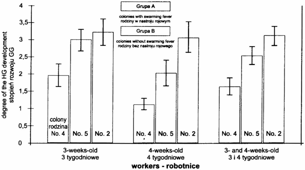
Figure 1 shows the average values for the degree of development of the HG in the workers classed by colony and age and originating from the samples
strona 28taken over the whole period of observation (colonies with swarming fever = densely punctured columns & colonies without swarming fever = column without punctation). About median values confidence intervals were graphically represented. The confidence intervals indicated that the degree of development of the HG was highly significantly (P<0.01) higher in the colonies with the swarming fever (colonies 2 and 5) than in the colonies without the swarming fever (colony 4). The development of the HG in 3-week-old workers coming from swarming colonies did not differ statistically (P<0.01). In 4-week-old workers of the colonies the difference was statistically highly significant (P<0.01). Colony 2 showed higher development of the HG than colony 5. Besides graphical representation, the differences were tested by analysis of variance, which confirmed that the differences were very highly significant (P<0.001). The method of subsequent testing also called the method of minimum differences confirmed that all the differences between the colonies were very highly significant.
The most important statistical characteristics are the values of the median and the mode (table 1). These characteristics exhibit maximum values in colonies with the swarming fever (colonies 2 and 5) and minimum values in colonies without the swarming fever (colonies 4).
Table 1 Median and mode of HG development in honeybee workers in 1997 – Mediana i średnia modalna stopnia rozwoju gruczołów gardzielowych robotnic w 1997r.
| workers > / robotnice; colony > / rodzina | 3-weeks-old (3 tygodniowe) No. 2 | 3-weeks-old No. 4 | 3-weeks-old No. 5 | 4-weeks-old (4 tygodniowe) No. 2 | 4-weeks-old No. 4 | 4-weeks-old No. 5 | 3- and 4-weeks-old (3 i 4 tygodniowe) No. 2 | 3- and 4-weeks-old No. 4 | 3- and 4-weeks-old No. 5 |
|---|---|---|---|---|---|---|---|---|---|
| median / rodzina mediana | 4 | 2 | 3 | 3,5 | 1 | 2 | 3,5 | 1 | 2,5 |
| mode / śr. modalna | 4 | 1 | 4 | 4 | 1 | 2 | 4 | 1 | 2 |
In 1998 there were also very good conditions for the rapid development of colonies. Based on the experience from the year 1997 the colonies were not stimulated to swarm. Instead, the following control measures were taken on the following dates:
- Colony 1 26 May honey harvest
- Colony 2 19 May inserting of 1 comb foundation / 26 May and 16 June honey harvest
- Colony 3 19 May inserting of 1 comb foundation / 16 June honey harvest
- Colony 4 12 May inserting of 2 comb foundations / 16 June honey harvest
- Colony 5 12 May addition of half-chamber and inserting of 2 comb foundations / 16 June honey harvest
Thus, all five colonies were prevented from premature swarming, but the development of the HG in 4-week-old workers among the colonies was not so different like in the year 1997 or in the experiments of the years 1993 and 1994 (Přidal, Háslbachová & Kubišová, 1996). Queen cells with eggs were only found in colony 5. A total of 789 workers were dissected which is 39 workers more than planned. In two years a total of 1102 workers were dissected.
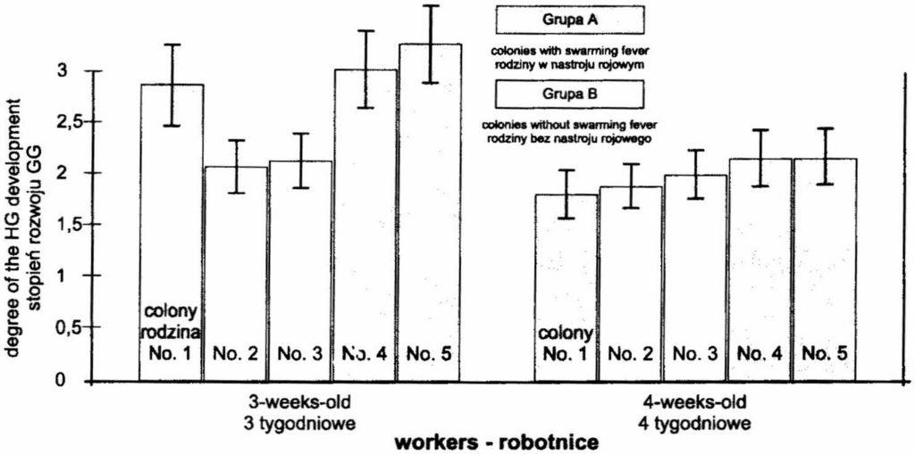
Figure 2 shows the average values for the development of the HG in 1998. The procedure is the same as in Figure 1 for the year 1997. The graphically represented confidence intervals illustrate highly significantly (P<0.01) higher development of the HG in 3-week-old workers of colonies 1, 4 and 5 (Group A) than in colonies 2 and 3 (Group B). In 4-week-old workers the differences were insignificant.
Like in the year 1997 in both cases analysis of variance and subsequent testing were made. Again, the differences were significant and in the group of 3-week-old workers the differences were very highly significant (P<0.001). Between the groups of 4-week-old workers the differences were markedly lower and the level of significance was P<0.05. Nonetheless, the graphically represented confidence intervals (Figure 2) did not confirm the differences and for this reason they are considered statistically insignificant.
Similarly like in 1997 the most transparent were the values of the median and the mode (table 2). These characteristics reached higher values in colonies of a group with the swarming fever and the lowest values were reported for groups without the swarming fever. The differences, however, are smaller than those found in the year 1997.
strona 30Table 2 Median and mode of HG development in honeybee workers in 1998 – Mediana i œrednia modalna stopnia rozwoju gruczo³ów gardzielowych robotnic
| workers > / robotnice; colony > / rodzina | 3-weeks-old – 3 tygodniowe No. 1 | No. 2 | No. 3 | No. 4 | No. 5 | 4-weeks-old – 4 tygodniowe No. 1 | No. 2 | No. 3 | No. 4 | No. 5 |
|---|---|---|---|---|---|---|---|---|---|---|
| median / rodzina mediana | 2 | 1,5 | 1,75 | 2 | 2 | 1 | 1 | 1,5 | 2 | 2 |
| mode / śr. modalna | 2 | 1 | 1 | 2 | 2 | 1 | 1 | 1 | 1 | 1 |
DISCUSSION
It is surprising that in both experimental years there was a high rate of the swarming fever. Kubišová & Háslbachová (1969) and Přidal, Háslbachová & Kubišová (1997) consistently reported that although the colonies were forced to swarm, not all of them did so and if they eventually swarmed, it was not until some time after the stimulation and not just at the beginning of the experiment. Under the conditions of high swarming rates it is difficult to maintain the planned number of workers in the samples collected. Moreover, prior to swarming fewer workers hatched and so it was difficult to have sufficient numbers of workers for marking.
In spite of the difficulties encountered the data obtained were not in conflict with the results from previous experiments (Přidal, Háslbachová & Kubišová, 1997). From the results it is evident that the colonies with the swarming fever had significantly higher developed HG, especially in 3-week-old workers, than the colonies that did not display the swarming fever. This was especially true of the year 1997 – colonies 2 and 5 showed the swarming fever and colony 4 did not. Somewhat unexpected is the statistically significant difference between the development of the HG in the 4-week-old workers coming from colonies 2 and 5 with the swarming fever. Colony 2 manifested greater development of the HG. This situation is explained logically by the results of the year (Kropáčová & Háslbachová, 1970 and Přidal, Háslbachová & Kubišová 1997). After swarming the queen in colony 2 did not lay eggs for a long time. This situation always causes changes in the development of the HG. The development is similar to that in a queenless colony. In such colonies the development of the HG and the ovaries at week 4 is high (degree III or IV). Another factor that could contribute to the difference is some of the treatments (adding foundation and later also half-chambers or also honey harvest) of the experimental colonies which were made to control premature swarming. The
strona 31last mentioned effect most probably contributed to the results of the year 1998. In that year there were only small differences in the development of the HG in 4-week-old workers. A preliminary explanation might be (Přidal, Háslbachová & Kubišová, 1997) that 3-week-old workers are a sort of physiological reserve in a bee colony that can easily respond to any changes in the colony.
For the first time the statistical characteristics of the median and the mode were also employed. They described the development of the HG more precisely than arithmetic mean. At early spring, the colonies, that will exhibit the swarming fever, always show the same decreasing of HG size as in colonies which will not swarm (Přidal, Háslbachová & Kubišová, 1997). Later, at the beginning of the swarming fever in colony (or queenless colony), decreasing of HG size is ceasing in 3-week and 4-week-old workers. Only at the time of full manifestation of the swarming fever all 3-week and 4-week-old workers show degrees III and mainly IV of development. This is why at the beginning of the swarming fever arithmetic mean characterises the changes in the development of the HG less markedly than at the end. The results obtained suggested that the values of the median and the mode were also higher in colonies with the swarming fever.
In 1998 only one colony swarmed (no. 5) and it showed all the characteristic changes in the development of the HG. These changes were also reported in colonies 1 and 4, which however failed to swarm. These colonies did not have eggs in queen cells, which is important according to the methodology for assessing the physiological status of a colony. A detailed analysis revealed that in colonies 1, 4, and 5 (Group A) there were dramatic fluctuations in brood rearing. There was most probably a dramatic increase in the area of the opened brood. And then only in these colonies a large number of marked bees aged 5 and 6 weeks were repeatedly reported. This phenomenon has never been found before in the colonies that exhibited uniform growth without swarming. This was typical of swarming colonies or queen-less colonies (Přidal, Háslbachová and Kubišová, 1997). It is, therefore, most probable that colonies 1 and 4 were committed to swarming and were just on the point of swarming fever exhibition – i.e. eggs in queen cells. Nonetheless, the colonies that did not swarm in the year 1998, i. e. colonies 2 and 3 (Group B) did not make queen cells for future use at all. However, the colonies of Group A did so very intensively and at control checks they had more than 15 queen cells in the brood-chamber, though they were not always with eggs. The building (presence) of queen cells in the colony has always been considered to have different importance. Our results, supported by dissection, confirmed that the presence of queen cells in large numbers can be an indication of the preparation for swarming or the period called the swarming fever (or mood).
strona 32The results of the dissections of the workers from colonies 1 and 4 that did not swarm indicated that these were colonies ready to swarm. This could mean that the difference in the development of the HG is, for example, genetically controlled. Nevertheless these colonies also showed other traits typical of colonies with the swarming fever – sudden changes in the rate of brood rearing and older workers, as mentioned above. The last mentioned trait must be further experimentally tested. It is still not clear if longevity is in this case induced also by physiological processes connected with swarming or chiefly by the genetic disposition of the colony, which was already proved (Kulincevic & Rothenbuhler, 1982).
SUMMARY
- It was repeatedly confirmed that predominantly 3-week-old workers coming from the colonies with the swarming fever have higher-developed HG than workers of the same age coming from the colonies without the swarming fever.
- The older workers (i.e. 5 and 6 weeks old) were recorded only in colonies containing workers which had highly developed HG also in their age of 3- and 4- weeks. In colonies without the swarming fever these older workers (more than 4 weeks of age) were not found.
- Only the colonies that exhibited the signs of preparedness for swarming, the swarming fever, (the high development of HG also in 3- and 4-week-old workers, the presence of older workers, etc.) built queen cells for future use. The colonies that did not show the swarming fever failed to do so.
ACKNOWLEDGEMENTS
We would like to thank to Prof. Dr. Sylvie Kubišová for help with interpretation of the results.
This work is dedicated to the seventieth anniversary of Ing. Hana Háslbachová, Ph.D. who worked as a scientist and a teacher of apidology at Department of Zoology and Apidology of Mendel University.
REFERENCES
Hess G. (1942)- Über den Einfluss der Weisellosigkeit und des Fruchtbarkeitsvitamins E auf die Ovarien der Bienenarbeiterin. Beihefte zur Schweiz. Bienen-Ztg, 1(2):33-110.
Kropáčová S., Háslbachová H. (1969)- The development of ovaries in worker honeybees in a queenright colony. J. Apic. Res. 8:57-64.
strona 33Kropáčová S., Háslbachová H. (1970)- The development of ovaries in worker honeybees in queenright colonies examined before and after swarming. J. Apic. Res. 9:65-70.
Kubišová S., Háslbachová H. (1985)- Studie o závislosti rozvoje vaječníků včel dělnic na stavu jejich hltanových žláz. [Study on dependence between the development of honeybee ovaries and phyryngeal glands]. Acta Univ. Agric. – Fac. agric. 33(4):253-258.
Kulincevic J.M., Rothenbuhler W.C. (1982)- Selection for length of life in the honey bee (Apis mellifera). Apidologie 13:347-352.
Lengefelder G. (1982)- Die Betrachtungen um Lebenführung des Bienenvolkes in Schwarmzeit. Imkerfreund 37: 308-314.
Přidal A., Háslbachová H., Kubišová S. (1997)- Stav hltanových žláz a vaječníků dělnic včely medonosné (Apis mellifera L.) v období rozvoje včelstev a rojení. [Condition of hypopharyngeal glands and ovaries in honeybee workers (Apis mellifera L.) during growth and swarming of colonies]. Acta Univ. Agric. Silvic. Mendel. Brun. (Brno) 45 (3-4): 51-58.
ROZWÓJ GRUCZOŁÓW GARDZIELOWYCH U ROBOTNIC PSZCZOŁY MIODNEJ W CZASIE WZROSTU OKRESU ROJOWEGO RODZIN PSZCZELICH
Přidal A., Šustek D.
Streszczenie
Rojenie się pszczół stanowi problem w pszczelarstwie, gdyż w niektórych latach trudno ograniczyć rójki w pasiekach do poziomu 10%. Zagadnienie nastroju rojowego i rojenia się pszczół nie zostało jeszcze do końca wyjaśnione. Celem przedstawionych tu badań było zbadanie rozwoju gruczołów gardzielowych (GG) w okresie wzrostu rodziny pszczelej i porównanie tej cechy u rodzin będących i nie będących w nastroju rojowym.
Badania prowadzono od 29 kwietnia do 26 czerwca 1997r. i od 28 kwietnia do 25 czerwca 1998 r., w 5 rodzinach pszczelich każdego roku. Podjęto specjalne działania dla wywołania nastroju rojowego, np. nie poszerzano gniazd, rodziny nie miały możliwości budowania plastrów itp. Co tydzień około 300 znakowanych pszczół, które wygryzły się w termostacie wpuszczano na powrót do rodziny macierzystej. Każdego tygodnia pszczoły znakowane były innym kolorem, co pozwalało rozróżnić wiek poszczególnych serii osobników. Próbki liczące 15 robotnic w wieku 3 i 4 tygodni pobierano z rodzin każdego tygodnia, utrwalano je w płynie Carnoa i umieszczano w 75% alkoholu do czasu wypreparowania gruczołów gardzielowych (GG). Notowano wszystkie zabiegi wykonane w rodzinach, ilość otwartego i zasklepionego czerwiu i biologiczny stan rodzin.
Wyniki badań potwierdziły, że robotnice 3-tygodniowe z rodzin w nastroju rojowym miały silniej rozwinięte GG niż robotnice tego samego wieku z rodzin bez nastroju rojowego. Starsze robotnice (tj. 5 i 6- tygodniowe) spotykano tylko w tych rodzinach, w których robotnice 3 i 4 -tygodniowe miały silnie rozwinięte GG. W rodzinach nie będących w nastroju rojowym nie spotykano robotnic starszych niż 4-tygodniowe. Tylko te
strona 34rodziny, w których występowały objawy przygotowania do rójki ( znaczny rozwój GG u pszczół 3 i 4-tygodniowych, obecność starszych robotnic etc.) budowały liczne miseczki matecznikowe. Rodziny nie wykazujące takich objawów nastroju rojowego nie czyniły tego. Uzyskane wyniki dyskutuje się w kontekście z innymi, wcześniejszymi badaniami i hipotezami.
Słowa kluczowe: Apis mellifera carnica, gruczoły gardzielowe, rojenie się pszczół.
Errata — błędy druku znalezione podczas digitalizacji
- s. 30, tab. 2: „œrednia” → średnia
- s. 30, tab. 2: „gruczo³ów” → gruczołów
- s. 33, piśmiennictwo: „phyryngeal” → pharyngeal
- s. 33, piśmiennictwo: „Lebenführung” → Lebensführung