

THE BULBUS GLAND OF DRONES (APIS MELLIFERA L)
tekst: Gudrun Koeniger, Heinz Hänel
Pszczelnicze Zeszyty Naukowe, Rok XL, 1996 — Gudrun Koeniger, Heinz Hänel
strona 45PSZCZELNICZE ZESZYTY NAUKOWE Rok XL, Nr 2 1996
Gudrun Koeniger, Heinz Hänel
Institut für Bienenkunde (Polytechnische Gesellschaft), Fachbereich Biologie der J.W. Goethe-Universität Frankfurt a.M., Karl-von-Frisch-Weg 2, 61440 Oberursel, GERMANY
Summary
In both newly emerged and 3,5 day old drones, the dorsal part of the bulbus epithelium consists of hypertrophied epidermal cells forming an epithelial gland. The cells above the chitin plates are columnar with a length up to 300 μm; at the sides of the bulbus they become shorter. The nucleus is situated in the basal part and has several nucleoli. Underneath the gland cells, the cuticle structure is modified to a cuticular membrane of variable thickness which contains numerous droplets of secretion. Two dorsal and two lateral chitin plates are inserted between the membrane and a ventral intima. They consist of densely packed globules of sclerotized cuticular material. The dorsal plates contain many pores and granules can be seen to migrate through the pores into the intima. The intima fills the lumen between the plates and in newly emerged drones it contains a supporting structure of filaments and only few droplets of secretion. At the age of 3,5 days more granules are enclosed, in sexually mature drones however, the secretion has merged and fills most of the intima. No gland cells could be found in the sections of old drones. The activity of the bulbus gland apparently is related to the maturation process of the drone.
Key words: Apis mellifera, drone, endophallus, bulbus gland, morphology
INTRODUCTION
The functional penis of the drone of all Apis species is a highly developed endophallic structure consisting of an eversible membrane. In contrast to endophallic tube of the simple adaegus of many other holometabolic insects The drone’s endophallus is greatly elaborated and differentiated into three major parts with various different structures and appendices. The vestibulum starts at the opening from the phallotreme. The middle part, called the cervix, opens into the bulbus, a large terminal expansion of the endophallus from which the ejaculatory duct originates (fig 1).
In Apis mellifera, the most obvious parts of the bulbus are the 2 chitinous plates, which lay at the dorsal side below the wall. They are separated by a narrow median external groove, from the posterior part of each plate a small triangular lateral plate with a spiny margin branches off. Ventral of the plates there is a “deep, brownish, semitransparent thickening of the cuticular intima which is extended as a pair of thin plates into the
strona 46lateral wall of the bulb. Externally the plates are covered by the thin epithelium of the bulb wall“ (Snodgrass, 1956). The inner walls of the bulb are entirely smooth (fig. 1a, b).
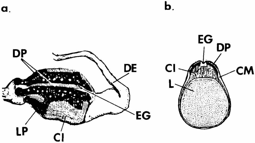
Woyke (1958) differentiates the wall of the bulbus into a thick and a thin part. The thick walled parts consist of a columnar epithelium which lays dorsally of the chitin plates. Woyke also describes the intima but calls it ‘connective substance’. Further, he describes additional columnar epithelium cells at the opening of the ejaculatory duct. In the thinly walled part of the bulb the epithelium is flat.
To clarify these contradictory descriptions of the bulbus’ epithelium we wanted to reexaminate and to explore whether or not a change occurs during the sexual maturation of the drone as known for other glandular epithelia (Bishop 1920).
MATERIAL AND METHODS
Sealed drone brood was put into an incubator (34,5°C, 75% humidity). Every 12 h the emerged drones were marked individually and introduced into a bee colony. They were recaptured at ages of 12 hours, 3-4 and 14 days. They were fixed in 10% formaldehyde and embedded either in
strona 47paraffin or for frozen sections in gelatine. They were stained with hematoxyline/eosin or in case of frozen sections with hematoxyline and fat red 7B by Merck.
RESULTS
- Sexually matured drone (age: 14 days)
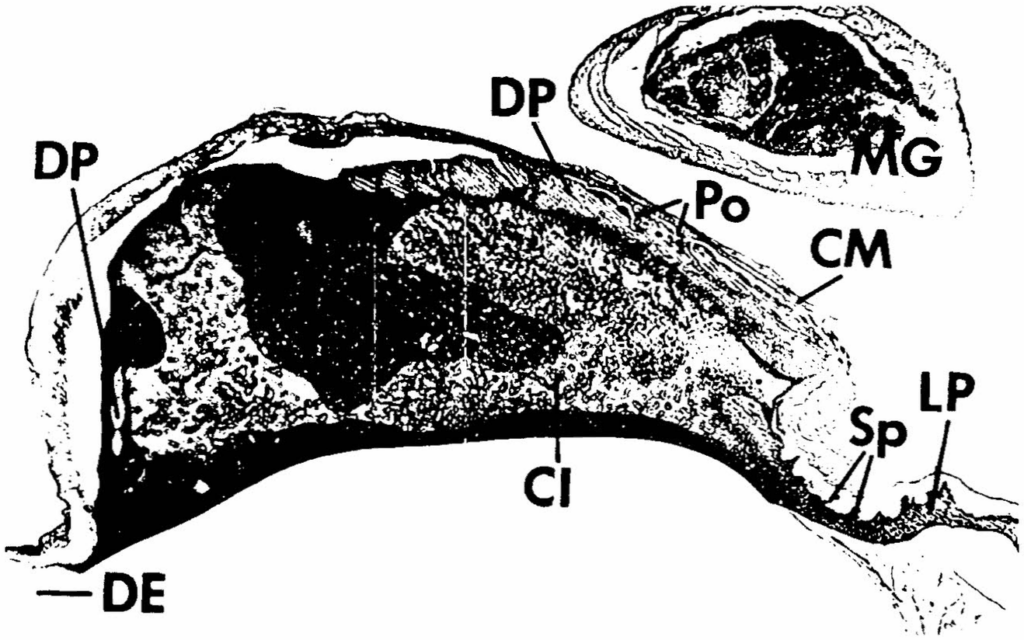
The longitudinal section of the bulbus (fig. 2) shows a lateral part of the bulubs, since the yellow lateral plates (LP) with its spiny margin (Sp) can be recognised easily. The dorsal plate (DP), also yellow, does not cover the cuticular intima (CI) continuously but is interrupted (fig. 1). The most conspicuous part in this section is the cuticular intima (CI), which appears brightly pink after staining. It consists mostly of a homogenous substance. It is well delimited by a membrane from the bulb’s lumen. The few holes (H) can be interpreted as channels. The lateral and dorsal chitin plates are firmly connected to the intima. The dorsal plates (DP) are penetrated by some pores (Po), while the lateral plates (LP) have tiny spines (Sp). Above the plates the bulbus wall is thin and contains only the remainders of epithelial cells. Only at the origin of the ejaculatory duct (ED) columnar epithelium cells are apparent. Also in the adjacent sections of this drone no epithelia cells could be found
- Young drones (age: 3-4 days)
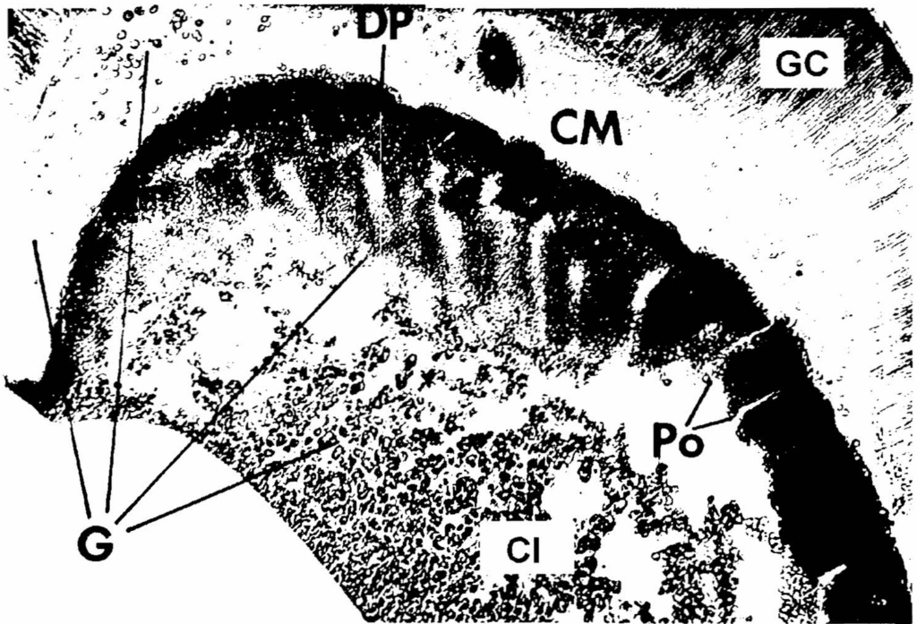
The longitudinal sections of both drones were conducted near the median of the bulb, only the dorsal plates (DP) can be seen (fig 3). In these younger drones, the wall above the plates is about 500μ thick. It consists of epithelial gland cells (GC), each cell about 300μ long, and a cuticular membrane (CM) in which some granules (G) are enclosed. The plates and the intima build another layer with a dimension up to 600 μm in the central part. In the frozen section (fig 3), the dorsal plates appear darker than the intima because the secretion is not stained. In contrast to the cuticular membrane, the structure of the plates does not consist of parallel layers but consists of many tiny round cuticular globules which are sclerotized and tanned. The plates are penetrated by many pores. Because of this structure, the plates are very fraile and often parts broke off during the dissection process. Tiny granules are attached at the dorsal and at the ventral side of the dorsal plates; some can be recognised at the opening of the pores and within the channel. Below the plates, they are arranged densely, except underneath the pores, where they are separated by a tubel-like space. Further down in the intima the granules are bigger and scattered. Few
strona 49granules have merged there. In the parts without secretion, a framework of thin filaments can be recognised.
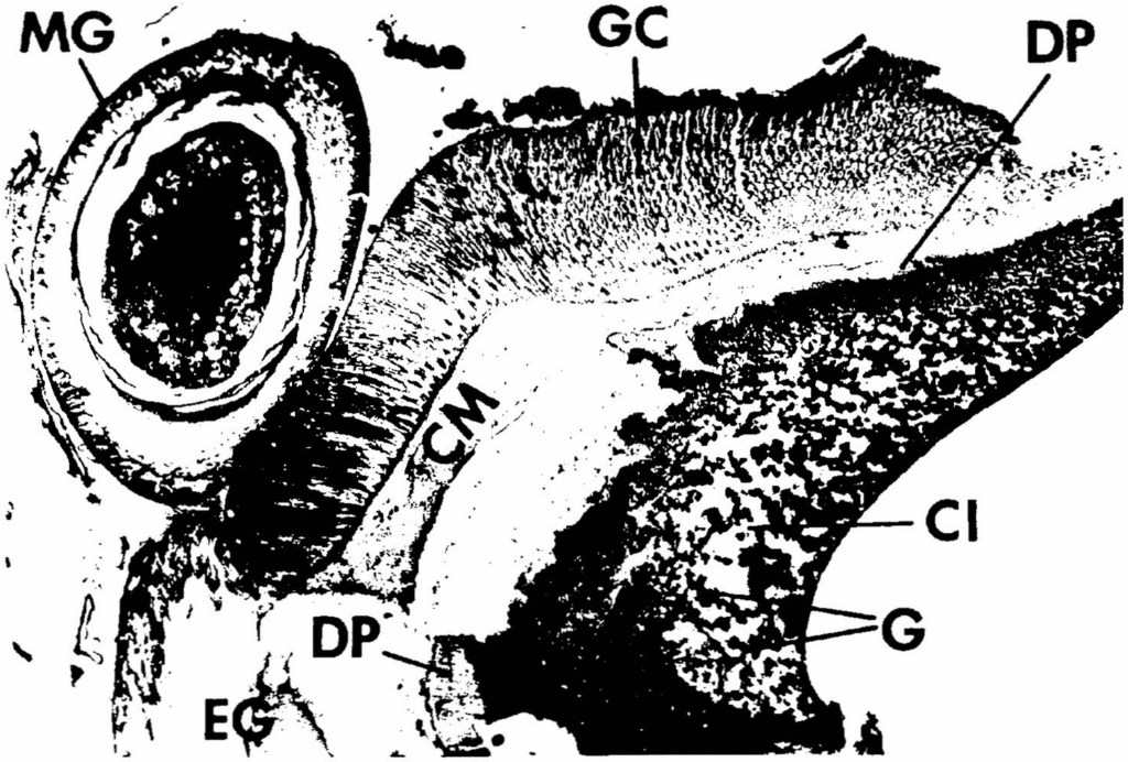
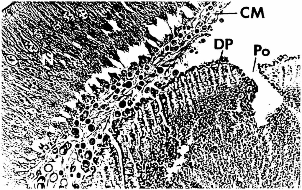
In the section of another drone (fig.4) the gland cells and the dorsal plate end at the cranial part of the bulbus well surrounded by the cuticular membrane. No lateral plates can be seen. In neighbouring sections of the same drone the space in front of the gland cells is filled by a cell complex which inflect from the external groove in a drop-shaped form. Droplets of secretion can be seen in the cuticular membrane and in the intima. Also in this section, the distribution is uneven underneath the plates, granules are small and densely packed and, further, ventral granules are bigger and less concentrated.
strona 50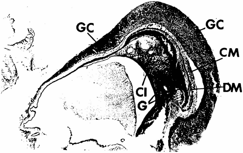
- Newly emerged drones (age: up to 12 hours)
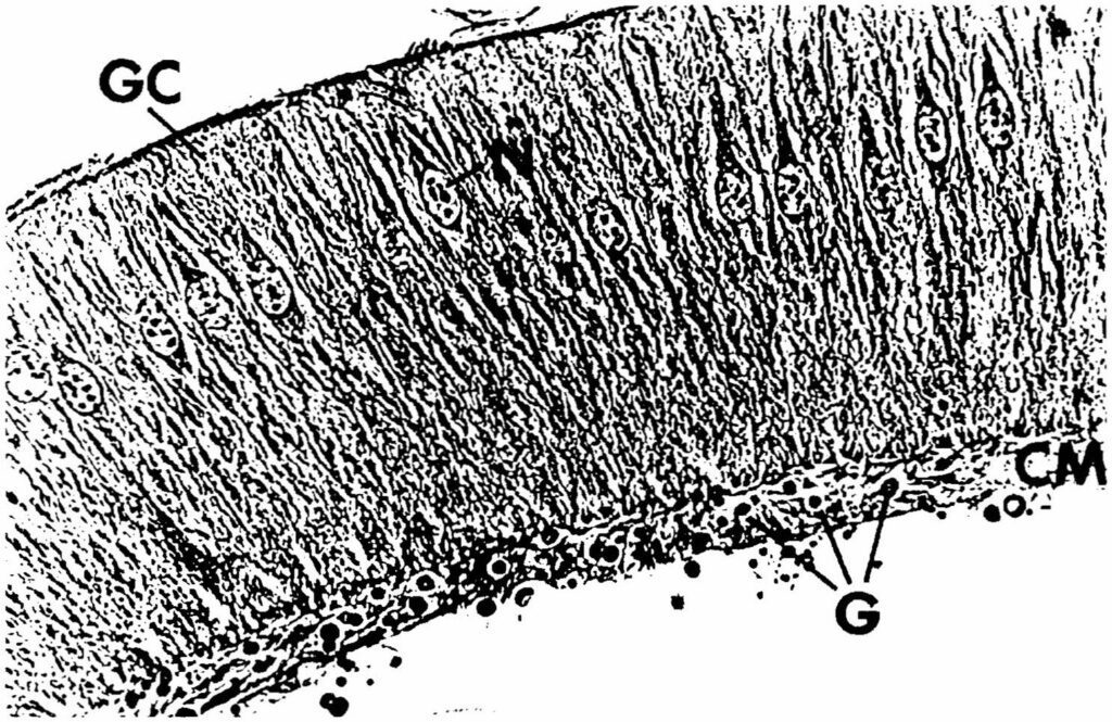
In figure 5 the intima of the bulb is only scarcely filled with granules. The supporting framework with filaments of different strength can be recognised which may be considered as part of the cuticular layer. Caudal and below the plates the secretory droplets (stained pink) are densely packed, in a transition zone they seem to change over into the chitin plates which are distinct by their yellow colour at the dorsal side. Numerous granules of secretion are in the cuticular membrane above the plates. The membrane surrounds all the bulbus but the granules can be found only beneath the gland cells. The longest gland cells (about 300 μm) are above the dorsal plates. At the sides, they become shorter until they transform into a normal flat epithelium. Along with the smaller gland cells, follows a thin projection of the intima. The nucleus of the gland cells is large and is located in the basal part of the cell, and several nucleoli are visible. In some parts the cells are compact columnar cells (fig. 7), in other parts they are goblet shaped (fig.6).
DISCUSSION
In our studies 14 day old drones showed few small epithelium cells on the dorsal side while in newly emerged drones the epithelium consists of large, active columnar gland cells. The different description of the bulbus wall of Snodgrass (1956) and Woyke (1958) seems to be based on the different age of the drones which they dissected. The epithelium in 14 day old drones has been either detached during the process of dissection or the cells have degenerated during maturation. Because of some degenerated cells left over at the cuticular membrane and the results of Snodgrass the latter hypothesis seems to be the most probable one.
Snodgrass (1956) interprets the complex of cuticular membrane, chitinous plates and intima as cuticular origin. The intima of newly emerged drones shows mainly a supporting framework of filaments, into which during sexual maturation granules are successively enclosed. In old drones the filaments cannot be seen in the light microscope, probably they are covered by the secretion. The lateral and dorsal plates are already present in emerging drones, also they are already tanned and scerotized. There structure of numerous little globules is different to the cuticular membrane. The membrane borders the gland cells and contains already in 1 day old drones some granules. In these gland cells, the nucleus is rather large. Together with the absence of fatty tissue, this can be considered as a sign of high metabolic and secretory activity. The situation in the cornual gland (Koeniger et al., 1996) is different where fatty tissue was found in the proximity of the secretory cells which seem to facilitate the metabolic process.
As the endophallus develops ontogenetically by inversion of the ectoderm (Zander 1922), the bulbus gland cells can be interpreted as an epidermal gland complex which secretes to the cuticle complex. The granules reach the intima passing first through the thin cuticular membrane and then through the pores of the chitinous plates. The intima is extended as a pair of thin plates along the lateral wall of the bulb (Snodgrass 1956, Woyke 1958)
The active secretions of the bulbus gland cells occur after emerging. This is coincides with several other processes which are connected to the maturation of drones. In the first days after emergence, spermatozoa migrate from the testes to the vesicula seminales where they undergo a stage of capacitation (Bishop 1920). The formation of mucus in the mucus glands also does not begin until after emergence of the drones (Bishop 1920. Mindt 1962). Also, the cornual gland secretion is produced only after emergence of the drone (Koeniger et al. 1990, 1996).
No research has been done on the chemical nature of the bulbus’ secretion nor about the biological function to our knowledge. The chitin
strona 53plates with the intima (connective substance plus reinforcing layer, Woyke 1958b, bow of the bulb Woyke and Ruttner 1958) become part of the mating sign and form a ring which holds the mucus when it is introduced into the sting chamber of the queen during the mating process (Woyke 1958b, Woyke and Ruttner 1958).
Further histological studies with an electron microscope and chemical analysis of the secretion are necessary to learn more about the function of the bulbus gland and its secretion in the life of the drone.
Acknowledgement: We thank Christel Rau for valuable assistance in taking and developing the pictures
REFERENCES
Bishop G.H. (1920) – Fertilisation in the honeybee. I. The male sexual organs: their histological structure and physiological functioning. J. exp. Zool., 31: 226-267
Koeniger G., Wissel M., Herth W., (1990) – Cornual secretion on the endophallus of the honeybee drone (Apis mellifera L): structure and secretion Apidologie, 21: 185-191
Koeniger G., Hänel H., Wissel M., Herth W., (1996) – Cornual gland of the honeybee drone (Apis mellifera L): structure and secretion. Apidologie, 27: 145-156
Koschevnikov G (1891) – Zur Anatomie der männlichen Geschlechtsorgane der Honigbiene. Zoolog. Anz.,14: 393-396
Mindt B. (1962) – Untersuchungen über das Leben der Drohnen, insbesondere Ernährung und Geschlechtsreife. Z. f. Bienenforschung, 6: 9-33
Snodgrass R.E. (1956) – Anatomy of the Honey Bee. Comstock Publications, London
Woyke J. (1958a) – The histological structure of the reproductive organs of the drone. Poznanskie Towarzystwo Przyjaciol Nauk. 19: 3-50
Woyke J. (1958b) – The process of mating in the honeybee. Pszcz Zesz Nauk. 2: 1-42
Woyke J., Ruttner F. (1958) – An anatomical study of the mating process in the honeybee. Bee World 39: 3-18
Zander E. (1922) – Der Bau der Biene. E.Ulmer Stuttgart Legends
GRUCZOŁY CZĘŚCI BULWIASTEJ APARATU KOPULACYJNEGO TRUTNIA (APIS MELLIFERA)
G. Koeniger, H. Hänel
Streszczenie
Znamię weselne, które truteń pozostawia w komorze żądłowej matki składa się z kilku części. Płytki chitynowe tworzą się w czasie rozwoju ontogenetycznego. Natomiast śluz i pomarańczowa substancja wytwarzają się w czasie dojrzewania płciowego dorosłego trutnia.
Woyke (1958) podaje, że na zewnątrz chitynowych płytek części bulwiastej znajduje się
strona 54jednowarstwowy nabłonek cylindryczny. Mrożone i parafinowe skrawki części bulwiastej wykazują, że grubość,nabłonka wynosi nad płytkami 200-300 μm i spłaszcza się ku bokom. U trutni 3-5 dniowych, pojawiają się granulki wydzieliny w przezroczystej warstwie oskórka między nabłonkiem a płytkami chitynowymi jak również w porach samych płytek. Wielkie zgromadzenie granulek znajduje się poniżej płytek w podłożu grubości 200-400 um.
Wynika z tego, że nabłonek nad płytkami chitynowymi jest jednowarstwowym gruczołem części bulwiastej, który staje się aktywnym po wygryzieniu się trutnia. Skład chemiczny wydzieliny, jak i jej biologiczne znaczenie nie jest znane. Możliwe, że bierze ona udział w tworzeniu substancji spajającej znajdującej się między płytkami, a opisanej przez Woykego.
Słowa kluczowe: Apis mellifera, trutnie, aparat kopulacyjny, gruczoły częśći bulwiastej, morfologia
Errata — błędy druku znalezione podczas digitalizacji
- s. 45: „adaegus” → aedeagus
- s. 47: „bulubs” → bulbus
- s. 48: „fraile” → fragile
- s. 48: „tubel-like” → tube-like
- s. 49, fig. 4: „broken of” → broken off
- s. 50, fig. 6: „goblar” → goblet
- s. 52: „scerotized” → sclerotized
- s. 52: „There structure” → Their structure
- s. 54, słowa kluczowe: „częśći” → części