

CHANGES IN THE PHARYNGEAL GLANDS OF BEES DURING THE DEVELOPMENT OF COLONIES
tekst: S. Kropáčová, Hana Haslbachovà, J. Pavel
Pszczelnicze Zeszyty Naukowe, Rok XIV, 1970 — S. Kropáčová, H. Haslbachová, J. Pavel
strona 129PSZCZELNICZE ZESZYTY NAUKOWE ROK XIV, NR 1—2—3 GRUDZIEŃ 1970
S. Kropáčová, H. Haslbachová, and J. Pavel
University of Agriculture, Brno, Czechoslovakia
INTRODUCTION
Already in 1927 the function of the pharyngeal glands of the honey-bee was elucidated (Soudek, 1927), consisting in the formation of royal jelly, i.e. the substance with which the young bees feed the just hatched out brood till the age of 3 days, further the queen’s larvae until pupation, as well as the egg-laying queen continuously. The application of royal jelly in human medicine has increased the general interest in the investigation of the factors linked with its production. General observations were already performed by Soudek (1927) who found that most bees collected on the brood had fully developed pharyngeal glands, while foraging bees, for the most part, had empty or dried up glands. Said investigator compiled a scale for the development of pharyngeal glands according to the shape and the size of the acini and demonstrated also that the activity of pharyngeal glands depended on the uptake of pollen in the food. Other authors continued their work and compared the development of pharyngeal glands on the basis of morphological characters with the function of bees in the colony, with the season of the year, with feeding bees, etc. (Lothmar, 1939; Wahl, 1959, and others).
Another method for the observation of the changes in the development of pharyngeal glands of bees were chemical analyses going in different directions. Of the existing very meagre studies on bee pharyngeal gland proteins attention is deserved, first and foremost, by the papers of Halberstadt (1966, 1967). Said author characterized the proteins of these glands with the aid of acrylamide gel electrophoresis in summer and winter bees and in those kept in little cages and found a number of typical electropherograms for worker bees of varying age and for drones. However, he did not occupy himself with their further characterization and
strona 130identification. Among further studies are of importance those which deal with the enzymatic properties of secretion proteins in young bees. Kratky (1931) confirmend the presence of invertase and amylase activities, intensively studied by Maurizio (1959, 1962, 1965); Ammon and Zoch (1957) demonstrated the activity of acid phosphatase, cholerinase, and other enzymes.
The present paper, continuing research on the basis of the existing knowledge, is focusing on the question of the electrophoretically differentiated structure of proteins in the bee pharyngeal gland with regard to the various developmental stages of the bee colony; furthermore it deals with the relationship between the development of pharyngeal glands and that of the bee ovaries, characterized in both cases by the morphological changes in the same developmental stage of the bee colony.
METHODS AND MATERIAL
Our observations were made on 14-day-old bees from two colonies and after their swarming out also from two swarms. Beginning with May 22nd and ending with July 17th, 1—2 days old marked bees were added every fortnight to the experimental colonies. For particulars see the paper written by Kropáčová and Haslbachová (1969).
A sample group of 30 bees taken on the 14th day of their life from each of the experimental colonies and swarms was subjected to carbon dioxide narcosis, where upon the bees were decapitated and their pharyngeal glands immediately excised. Under the microscope the size and the shape of the individual gland acini of each experimental bee were recorded and classified according to Soudek’s scale (1927) into 4 groups. The glands were then washed in distilled water and separated with the aid of an ultrafilter from the aqueous medium. From the ultrafilter they were collected into a cuvette containing 0.15 ml 1 M saccharose solution, homogenized, the supernatant being put in a starch block for electrophoresis. The time of run was 3.5 hours a discontinuity TRIS — citrate-borate buffer of pH 8.6 and a voltage of 440 V being used. Protein detection was performed with the aid of a 10 B amidoblack solution.
For determining the correlation between the development of the ovaries and the pharyngeal glands of bees, the developmental stage of the pharyngeal glands of each sample of 30 bees was converted to a system of points so that the first class of Soudek’s system, i.e. the most developed glands, was multiplied by the number n = 4, the second class by the number n — 1, and so on. The points referring to the development of the ovaries were taken from the paper written by Kropáčová and Haslbachová (1970).
strona 131RESULTS
The characteristic pictures of the electrophoretic distribution of proteins isolated from the pharyngeal glands of bee colonies in the various stages of development are recorded in Fig. 1. Taking a general view it can be said that in the pharyngeal gland a relatively heterogeneous pro-
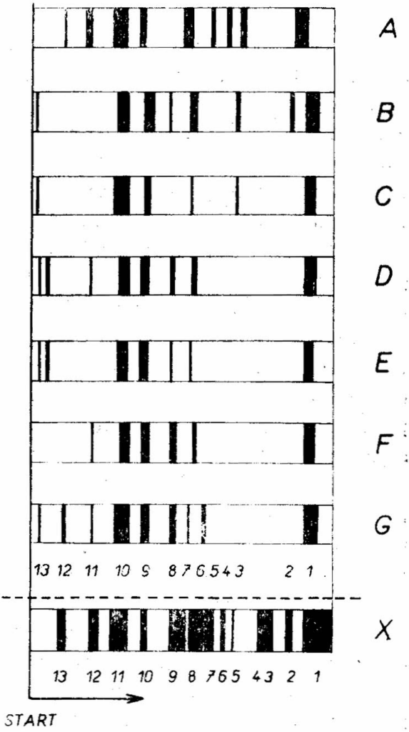
Fig. 1.
Scheme of electrophoretic distribution of main fractions of pharyngeal gland proteins in starch gel in the individual developmental stages of the colony:
Schemat elektroforetycznego rozdziału głównych frakcji białek z gruczołów gardzielowych w żelu skrobiowym w pojedynczych stadiach rozwoju rodziny pszczelej:
A Bee colony No. 2 two days before swarming rodzina nr 2 na 2 dni przed rójką
B Bee colony No. 2 twelve days after swarming out, queen not yet laying eggs rodzina nr 2 w 12 dni po wyjściu roju, matka jeszcze nieczerwiąca
C Swarm from colony No 2 twelve days after leaving the colony, queen laying eggs. rój z nr 2 w 12 dni po wyjściu, matka czerwiąca
D Bee colony No 2 twenty six days after swarming out, queen not yet laying eggs rodzina nr 2 w 26 dni po rójce, matka jeszcze nieczerwiąca
E Swarm from colony No 2 twenty six days after leaving the colony rój z nr 2 w 26 dni po wyjściu
F Bee colony No. 2 forty days after swarming out, queen not yet laying eggs rodzina nr 2 w 40 dni po rójce, matka jeszcze nieczerwiąca
G Swarm from bee colony No. 2 forty days after leaving the colony rój z nr 2 w 40 dni po wyjściu
X Typical electropherogram of pig blood serum proteins with identification of the main fractions
typowy elektroferogram białek z surowicy krwi świń z oznaczeniem frakcji podstawowych
1 albumin 2 postalbumins (α1) 3 Fα1 — globulins 4 β1 — globulins 5 β1 — globulins 6 ceruloplasmin 7 α2 — globulins 8 haptoglobin 9 β2 — transferins 10 amylase 11 Sα2 — globulins 12 Sα2 — globulins 13 low-density lipoproteins
strona 132tein complex is found, which during starch gel electrophoresis breaks up into a number of fractions of a hitherto unknown chemical character. To rely at least in the most general sense of orientation on some model, we argumented the mentioned picture in the last column by the illustration of the electrophoretic distribution of proteins in the pig blood serum with a description of their main fractions (Pavel and Harásek, 1971). If we perform a mutual comparison of the protein electropherograms of the pharyngeal glands taken from the bee colonies at various stages of their development, we can find the following two most typical facts: Significant changes in the structure of pharyngeal gland proteins are connected with the swarming process, where both in the swarmed out bee colony and in the swarm a significant reduction of the number of present fractions sets in, compared with the colony prior to swarming. This reduction is limited to the time until more intensive egg-laying by the queen occurs, when the worker bees begin to use the secretions of the pharyngeal glands in the nutrition of the brood. In that period a certain multiplication of the protein spectrum gradually occurs again, as can be seen in the electropherogram marked G. As long as the queen is not able to lay eggs, the function of the pharyngeal glands of bees stagnates and the number of the present fractions remains low (electropherogram F). Besides these phenomena we can also count with a certain seasonal variability in the structure of pharyngeal gland proteins.
For completing this picture we added still a photograph of a starch block with distributed samples of pharyngeal gland proteins from bee colonies of that period, partly in cases where the queen is laying eggs, partly where she is not laying eggs (Fig. 2).
As to the actual characterization of pharyngeal gland proteins no reliable answer can be given as yet. As has already been said, we added a pig blood serum protein distribution electropherogram for curiosity’s sake. From the relevant comparison it is to be seen that in pharyngeal gland proteins the mobility of the main fractions corresponds, for example, to the position of albumin (fraction 1), while the following two important fractions lie at the level of the amylase fraction and the pig blood serum slow globulin (Sα2) fraction, etc. It goes without saying that from what has been said no reciprocal identity can be deduced.
When comparing the individual electropherograms with the results of the morphological evaluation of the development of pharyngeal glands, no correlation between the quantity and the quality of secretion protein fractions and the developmental stage of the glands can be found. This finding is in agreement with that of Halberstadt (1966).
So much the more did we take interest in the relationship between the development of the pharyngeal glands and the ovaries of these bees. Practical bee-keeping, in fact, assumes that a better development of the worker bee ovaries in the period, when in the bee colony there is a little
strona 133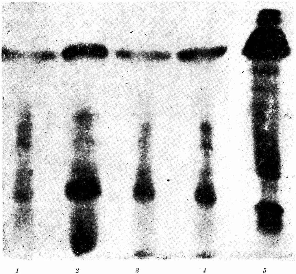
[Fotografia bloku skrobiowego — pięć ścieżek elektroforetycznych, bez tekstu; pod ścieżkami numery: 1 2 3 4 5.]
Fig. 2.
Photograph of starch block with the individual behaviours of the analyzed pharyngeal gland samples:
Fotografia wyciągu skrobiowego z wyodrębnionych próbek analizowanych gruczołów gardzielowych:
1 electropherogram of bee colony forty days after swarming, queen not yet laying eggs elektroferogram rodziny w 40 dni po rójce, matka nieczerwiąca
2 swarm from the same bee colony forty days after swarming, queen laying eggs rój z tejże rodziny w 40 dni po wyjściu, matka czerwiąca
3 the other bee colony under study, forty four days after swarming, queen laying eggs inna rodzina w badaniu w 44 dni po rójce, matka czerwiąca
4 swarm from preceding bee colony forty four days after swarming, queen laying eggs rój z tejże rodziny w 44 dni po wyjściu, matka czerwiąca
5 electropherogram of pig blood serum proteins used for comparison elektroferogram białek z surowicy krwi świń dla porównania
strona 134unsealed brood, is connected with the very consumption of excess royal jelly produced by these bees.
In the comparison of the point values of both factors, a positive correlation in both swarms and in the bee colony, where the young queen begins to lay eggs soon after swarming out (r = +0.649, r = +0.941, resp., P<0.05 for both evaluations) was demonstrated. In the bee colony where the queen had not laid eggs for a long time, this correlation was not confirmed; on the contrary, the trend of an opposite correlation appeared, where with the decrease of hypertrophy of the pharyngeal glands the ovaries developed more. The correlation, however, was not statistically significant (r = —0.553). On the basis of the results of these experiments it can be said that the influence of the excess production of royal jelly by young bees on the development of their ovaries cannot be unambiguously confirmed.
SUMMARY
The changes of proteins isolated from the pharyngeal gland of the bee, tested for their electrophoretic distribution in starch gel, in various stages of development are described. The main changes are recorded partly in the period of swarming, partly in a later period when young queens are present in the bee colonies. Furthermore it is possible to say that the electrophoretic mobility of the main fractions of pharyngeal gland proteins in comparison with the electropherogram of pig blood serum proteins corresponds to the position typical of albumin, serum amylase, and slow α2-globulins.
No correlation between the shape and the size of the pharyngeal gland acini and the quantity and the quality of the protein fractions in the secretions from these glands has been found.
In two groups of bee colonies a direct relationship between the developmental stage of the pharyngeal glands and that of the worker bee ovaries has been demonstrated.
REFERENCES
Ammon R., Zoch E.: Arzneimittelforschung 7, 699—702 (1957).
Halberstadt K.: Ann. Abeille 9, 153—163 (1966).
Halberstadt K.: Ann. Abeille 10, 119—132 (1967).
Kratky E.: Z. wissensch. Zool. 139, 120—200 (1931).
Kropáčova S., Haslbachová H.: J. Apic. Res. 8, 57—64 (1969).
Kropáčova S., Haslbachová H.: J. Apic. Res. in press. (1970).
Lothmar, R.: Schweiz. Bienenztg. 75, 262—265 (19[?]9).
Maurizio, A.: Bee World 40, 275—283 (1959)
strona 135Maurizio, A.: Ann. Abeille 5, 215—232 (1962).
Maurizio, A.: Ann. Abeille 8, 113—128 (1965).
†† Soudek, Š.: Bulletin de l’école superieure d’agronomie, Brno, Czechoslovakia, 10, 1—63 (1927).
Wahl, O.: Leipzig, Bienenztg: 73, 105—108 (1959).
†† Pavel, J., Harásek, E.: Přispěvek k poznáni heterogenity sérových bílkovin u prasat (A Contribution to the Knowledge of the Heterogeneity of Pig Serum Proteins) (prepared for press) (1971).
ZMIANY W GRUCZOŁACH GARDZIELOWYCH ROBOTNIC W OKRESIE ROZWOJU RODZIN PSZCZELICH
S. Kropáčova, H. Halsbachová J. Pavel,
Wyższa Szkoła Rolnicza, Brno CSRS
Zmiany protein, wyizolowanych z gruczołów gardzielowych pszczoły miodnej, badane przez rozdział elektroforetyczny w żelu skrobiowym — przeanalizowano w różnych stadiach rozwoju rodziny pszczelej.
Najpoważniejsze zmiany zaobserwowano w okresie rójki i później, kiedy w roju znajdowała się młoda matka. Można też mówić, że elektroforetyczna zmienność głównych frakcji protein z gruczołów gardzielowych pszczoły robotnicy w porównaniu z elektroforegramami surowicy krwi świń odpowiada typowemu położeniu albuminy, surowicy amylazy i wolnych α2-globulin.
Nie stwierdzono korelacji między kształtem i wielkością gruczołów gardzielowych a ilością i jakością frakcji bialkowych w wydzielinach tych gruczołów.
W dwu grupach rojów zaobserwowano u robotnic prostą zależność między stopniem rozwoju gruczołów gardzielowych i jajników.
ИЗМЕНЕНИЯ В ГЛОТОЧНЫХ ЖЕЛЕЗАХ РАБОТНИЦ В ПЕРИОДЕ РАЗВИТИЯ ПЧЕЛИНОЙ СЕМЬИ
С. Кропачова, Г. Галсбахова, Я. Павел
Резюме
Изменеия протеин, обособленных из глоточных желез медоносной пчелы в разных стадиях развития пчелиной семьи, исследовано разделяя их при помощи электрофорезы в крахмальном геле.
Самые сильные изменения наблюдались в периоде роения и после него, когда в семье была молодая матка. Можно сказать, что изменчивость главных фракций полученных при электрофорезе протеин из глоточных желез пчел работниц, при сравнении с электрофорезограммами сыворотки крови свиней, отвечает положению альбумин сыворотки: амилазы и свободных α2 — глобулин.
Не обнаружено корреляции между формой и величиной глоточных желез а количеством и качеством белковых фракций в секрете этих желез. В двух группах семей наблюдалось прямая зависимость между степенью развития у работниц глоточных желез и яичников.
Errata — błędy druku znalezione podczas digitalizacji
- s. 130: „confirmend” → confirmed
- s. 131, ryc. 1, legenda, poz. 9: „transferins” → transferrins
- s. 132: „argumented” → augmented
- s. 134–135, bibliografia i główka streszczenia polskiego, 3×: „Kropáčova” → Kropáčová
- s. 135, główka streszczenia polskiego: „Halsbachová” → Haslbachová
- s. 135, streszczenie polskie: „bialkowych” → białkowych
- s. 135, główka streszczenia rosyjskiego: „Галсбахова” → Гаслбахова
- s. 135, streszczenie rosyjskie: „Изменеия” → Изменения